You are browsing environment: HUMAN GUT
CAZyme Information: MGYG000001364_01536
You are here: Home > Sequence: MGYG000001364_01536
Basic Information |
Genomic context |
Full Sequence |
Enzyme annotations |
CAZy signature domains |
CDD domains |
CAZyme hits |
PDB hits |
Swiss-Prot hits |
SignalP and Lipop annotations |
TMHMM annotations
Basic Information help
| Species | Phocaeicola plebeius | |||||||||||
|---|---|---|---|---|---|---|---|---|---|---|---|---|
| Lineage | Bacteria; Bacteroidota; Bacteroidia; Bacteroidales; Bacteroidaceae; Phocaeicola; Phocaeicola plebeius | |||||||||||
| CAZyme ID | MGYG000001364_01536 | |||||||||||
| CAZy Family | GH16 | |||||||||||
| CAZyme Description | Beta-agarase | |||||||||||
| CAZyme Property |
|
|||||||||||
| Genome Property |
|
|||||||||||
| Gene Location | Start: 76329; End: 77279 Strand: - | |||||||||||
CAZyme Signature Domains help
| Family | Start | End | Evalue | family coverage |
|---|---|---|---|---|
| GH16 | 51 | 310 | 3.2e-111 | 0.9959349593495935 |
CDD Domains download full data without filtering help
| Cdd ID | Domain | E-Value | qStart | qEnd | sStart | sEnd | Domain Description |
|---|---|---|---|---|---|---|---|
| cd02178 | GH16_beta_agarase | 1.21e-89 | 28 | 310 | 4 | 257 | Beta-agarase, member of glycosyl hydrolase family 16. Beta-agarase is a glycosyl hydrolase family 16 (GH16) member that hydrolyzes the internal beta-1,4-linkage of agarose, a hydrophilic polysaccharide found in the cell wall of Rhodophyceaea, marine red algae. Agarose is a linear chain of galactose units linked by alternating L-alpha-1,3- and D-beta-1,4-linkages that are additionally modified by a 3,6-anhydro-bridge. Agarose forms thermo-reversible gels that are widely used in the food industry or as a laboratory medium. While beta-agarases are also found in two other families derived from the sequence-based classification of glycosyl hydrolases (GH50, and GH86) the GH16 members are most abundant. This domain adopts a curved beta-sandwich conformation, with a tunnel-shaped active site cavity, referred to as a jellyroll fold. |
| cd00413 | Glyco_hydrolase_16 | 5.69e-22 | 51 | 310 | 1 | 210 | glycosyl hydrolase family 16. The O-Glycosyl hydrolases are a widespread group of enzymes that hydrolyse the glycosidic bond between two or more carbohydrates, or between a carbohydrate and a non-carbohydrate moiety. A glycosyl hydrolase classification system based on sequence similarity has led to the definition of more than 95 different families inlcuding glycosyl hydrolase family 16. Family 16 includes lichenase, xyloglucan endotransglycosylase (XET), beta-agarase, kappa-carrageenase, endo-beta-1,3-glucanase, endo-beta-1,3-1,4-glucanase, and endo-beta-galactosidase, all of which have a conserved jelly roll fold with a deep active site channel harboring the catalytic residues. |
| cd08023 | GH16_laminarinase_like | 9.88e-13 | 50 | 310 | 2 | 235 | Laminarinase, member of the glycosyl hydrolase family 16. Laminarinase, also known as glucan endo-1,3-beta-D-glucosidase, is a glycosyl hydrolase family 16 member that hydrolyzes 1,3-beta-D-glucosidic linkages in 1,3-beta-D-glucans such as laminarins, curdlans, paramylons, and pachymans, with very limited action on mixed-link (1,3-1,4-)-beta-D-glucans. |
CAZyme Hits help
| Hit ID | E-Value | Query Start | Query End | Hit Start | Hit End |
|---|---|---|---|---|---|
| EDY95404.1 | 1.12e-248 | 1 | 316 | 1 | 316 |
| BBK89488.1 | 5.62e-154 | 38 | 316 | 40 | 322 |
| AXP07831.1 | 3.38e-139 | 23 | 316 | 25 | 323 |
| AXP07836.1 | 9.82e-125 | 26 | 310 | 20 | 289 |
| AIY13463.1 | 1.86e-116 | 19 | 314 | 46 | 330 |
PDB Hits download full data without filtering help
| Hit ID | E-Value | Query Start | Query End | Hit Start | Hit End | Description |
|---|---|---|---|---|---|---|
| 5T9X_A | 4.32e-155 | 38 | 316 | 61 | 343 | Crystalstructure of BuGH16Bwt [Bacteroides uniformis],5T9X_B Crystal structure of BuGH16Bwt [Bacteroides uniformis],5T9X_C Crystal structure of BuGH16Bwt [Bacteroides uniformis] |
| 3WZ1_A | 1.75e-118 | 28 | 314 | 4 | 276 | Catalyticdomain of beta-agarase from Microbulbifer thermotolerans JAMB-A94 [Microbulbifer thermotolerans] |
| 4ATF_A | 9.39e-110 | 25 | 315 | 10 | 307 | Crystalstructure of inactivated mutant beta-agarase B in complex with agaro-octaose [Zobellia galactanivorans],4ATF_B Crystal structure of inactivated mutant beta-agarase B in complex with agaro-octaose [Zobellia galactanivorans],4ATF_C Crystal structure of inactivated mutant beta-agarase B in complex with agaro-octaose [Zobellia galactanivorans],4ATF_D Crystal structure of inactivated mutant beta-agarase B in complex with agaro-octaose [Zobellia galactanivorans] |
| 1O4Z_A | 1.16e-109 | 25 | 315 | 41 | 338 | TheThree-dimensional Structure Of Beta-agarase B From Zobellia Galactanivorans [Zobellia galactanivorans],1O4Z_B The Three-dimensional Structure Of Beta-agarase B From Zobellia Galactanivorans [Zobellia galactanivorans],1O4Z_C The Three-dimensional Structure Of Beta-agarase B From Zobellia Galactanivorans [Zobellia galactanivorans],1O4Z_D The Three-dimensional Structure Of Beta-agarase B From Zobellia Galactanivorans [Zobellia galactanivorans] |
| 6AII_A | 1.11e-83 | 25 | 311 | 2 | 322 | Catalyticdomain of PdAgaC [Persicobacter] |
Swiss-Prot Hits download full data without filtering help
| Hit ID | E-Value | Query Start | Query End | Hit Start | Hit End | Description |
|---|---|---|---|---|---|---|
| B5CY73 | 2.24e-249 | 1 | 316 | 1 | 316 | Beta-agarase OS=Phocaeicola plebeius (strain DSM 17135 / JCM 12973 / M2) OX=484018 GN=BACPLE_01670 PE=1 SV=1 |
| Q9RGX8 | 8.02e-109 | 25 | 315 | 56 | 353 | Beta-agarase B OS=Zobellia galactanivorans (strain DSM 12802 / CCUG 47099 / CIP 106680 / NCIMB 13871 / Dsij) OX=63186 GN=agaB PE=1 SV=1 |
| D7GXG4 | 1.68e-82 | 1 | 310 | 1 | 373 | Beta-agarase D OS=Zobellia galactanivorans (strain DSM 12802 / CCUG 47099 / CIP 106680 / NCIMB 13871 / Dsij) OX=63186 GN=agaD PE=1 SV=1 |
| G0L322 | 2.41e-77 | 28 | 310 | 22 | 284 | Beta-agarase A OS=Zobellia galactanivorans (strain DSM 12802 / CCUG 47099 / CIP 106680 / NCIMB 13871 / Dsij) OX=63186 GN=agaA PE=1 SV=1 |
| A8W969 | 1.62e-53 | 12 | 310 | 8 | 296 | Beta-agarase AgaB34 OS=Agarivorans albus OX=182262 GN=agaB34 PE=3 SV=1 |
SignalP and Lipop Annotations help
This protein is predicted as SP
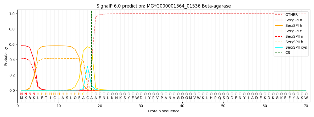
| Other | SP_Sec_SPI | LIPO_Sec_SPII | TAT_Tat_SPI | TATLIP_Sec_SPII | PILIN_Sec_SPIII |
|---|---|---|---|---|---|
| 0.000866 | 0.572409 | 0.426008 | 0.000309 | 0.000213 | 0.000191 |