You are browsing environment: HUMAN GUT
CAZyme Information: MGYG000001374_01580
You are here: Home > Sequence: MGYG000001374_01580
Basic Information |
Genomic context |
Full Sequence |
Enzyme annotations |
CAZy signature domains |
CDD domains |
CAZyme hits |
PDB hits |
Swiss-Prot hits |
SignalP and Lipop annotations |
TMHMM annotations
Basic Information help
| Species | Mediterraneibacter torques | |||||||||||
|---|---|---|---|---|---|---|---|---|---|---|---|---|
| Lineage | Bacteria; Firmicutes_A; Clostridia; Lachnospirales; Lachnospiraceae; Mediterraneibacter; Mediterraneibacter torques | |||||||||||
| CAZyme ID | MGYG000001374_01580 | |||||||||||
| CAZy Family | GH73 | |||||||||||
| CAZyme Description | hypothetical protein | |||||||||||
| CAZyme Property |
|
|||||||||||
| Genome Property |
|
|||||||||||
| Gene Location | Start: 697510; End: 699036 Strand: - | |||||||||||
CAZyme Signature Domains help
| Family | Start | End | Evalue | family coverage |
|---|---|---|---|---|
| GH73 | 213 | 345 | 1.5e-17 | 0.9765625 |
CDD Domains download full data without filtering help
| Cdd ID | Domain | E-Value | qStart | qEnd | sStart | sEnd | Domain Description |
|---|---|---|---|---|---|---|---|
| pfam01510 | Amidase_2 | 1.08e-35 | 23 | 154 | 1 | 121 | N-acetylmuramoyl-L-alanine amidase. This family includes zinc amidases that have N-acetylmuramoyl-L-alanine amidase activity EC:3.5.1.28. This enzyme domain cleaves the amide bond between N-acetylmuramoyl and L-amino acids in bacterial cell walls (preferentially: D-lactyl-L-Ala). The structure is known for the bacteriophage T7 structure and shows that two of the conserved histidines are zinc binding. |
| cd06583 | PGRP | 2.43e-27 | 23 | 155 | 1 | 126 | Peptidoglycan recognition proteins (PGRPs) are pattern recognition receptors that bind, and in certain cases, hydrolyze peptidoglycans (PGNs) of bacterial cell walls. PGRPs have been divided into three classes: short PGRPs (PGRP-S), that are small (20 kDa) extracellular proteins; intermediate PGRPs (PGRP-I) that are 40-45 kDa and are predicted to be transmembrane proteins; and long PGRPs (PGRP-L), up to 90 kDa, which may be either intracellular or transmembrane. Several structures of PGRPs are known in insects and mammals, some bound with substrates like Muramyl Tripeptide (MTP) or Tracheal Cytotoxin (TCT). The substrate binding site is conserved in PGRP-LCx, PGRP-LE, and PGRP-Ialpha proteins. This family includes Zn-dependent N-Acetylmuramoyl-L-alanine Amidase, EC:3.5.1.28. This enzyme cleaves the amide bond between N-acetylmuramoyl and L-amino acids, preferentially D-lactyl-L-Ala, in bacterial cell walls. The structure for the bacteriophage T7 lysozyme shows that two of the conserved histidines and a cysteine are zinc binding residues. Site-directed mutagenesis of T7 lysozyme indicates that two conserved residues, a Tyr and a Lys, are important for amidase activity. |
| smart00644 | Ami_2 | 2.15e-26 | 22 | 152 | 1 | 126 | Ami_2 domain. |
| COG3023 | AmpD | 9.87e-24 | 10 | 155 | 32 | 167 | N-acetyl-anhydromuramyl-L-alanine amidase AmpD [Cell wall/membrane/envelope biogenesis]. |
| COG5632 | CwlA | 7.66e-18 | 17 | 180 | 17 | 174 | N-acetylmuramoyl-L-alanine amidase CwlA [Cell wall/membrane/envelope biogenesis]. |
CAZyme Hits help
| Hit ID | E-Value | Query Start | Query End | Hit Start | Hit End |
|---|---|---|---|---|---|
| QMW80001.1 | 1.95e-109 | 5 | 506 | 2 | 500 |
| QIB57222.1 | 1.95e-109 | 5 | 506 | 2 | 500 |
| AFC63685.1 | 1.81e-106 | 6 | 508 | 4 | 499 |
| ADL53623.1 | 4.47e-101 | 47 | 506 | 47 | 487 |
| QHJ84983.1 | 5.19e-91 | 5 | 182 | 2 | 179 |
PDB Hits download full data without filtering help
| Hit ID | E-Value | Query Start | Query End | Hit Start | Hit End | Description |
|---|---|---|---|---|---|---|
| 2BH7_A | 4.86e-13 | 23 | 146 | 28 | 154 | Crystalstructure of a SeMet derivative of AmiD at 2.2 angstroms [Escherichia coli K-12] |
| 2WKX_A | 4.86e-13 | 23 | 146 | 28 | 154 | Crystalstructure of the native E. coli zinc amidase AmiD [Escherichia coli K-12],3D2Y_A Complex of the N-acetylmuramyl-L-alanine amidase AmiD from E.coli with the substrate anhydro-N-acetylmuramic acid-L-Ala-D-gamma-Glu-L-Lys [Escherichia coli str. K-12 substr. MG1655],3D2Z_A Complex of the N-acetylmuramyl-L-alanine amidase AmiD from E.coli with the product L-Ala-D-gamma-Glu-L-Lys [Escherichia coli str. K-12 substr. MG1655] |
| 1YB0_A | 9.18e-09 | 7 | 174 | 5 | 157 | Structureof PlyL [Bacillus anthracis],1YB0_B Structure of PlyL [Bacillus anthracis],1YB0_C Structure of PlyL [Bacillus anthracis] |
| 2L47_A | 1.40e-08 | 7 | 182 | 5 | 165 | Solutionstructure of the PlyG catalytic domain [Bacillus phage Gamma] |
| 2AR3_A | 5.88e-08 | 7 | 174 | 5 | 157 | ChainA, prophage lambdaba02, n-acetylmuramoyl-l-alanine amidase, family 2 [Bacillus anthracis],2AR3_B Chain B, prophage lambdaba02, n-acetylmuramoyl-l-alanine amidase, family 2 [Bacillus anthracis],2AR3_C Chain C, prophage lambdaba02, n-acetylmuramoyl-l-alanine amidase, family 2 [Bacillus anthracis] |
Swiss-Prot Hits download full data without filtering help
| Hit ID | E-Value | Query Start | Query End | Hit Start | Hit End | Description |
|---|---|---|---|---|---|---|
| O51481 | 1.42e-20 | 217 | 347 | 75 | 197 | Uncharacterized protein BB_0531 OS=Borreliella burgdorferi (strain ATCC 35210 / DSM 4680 / CIP 102532 / B31) OX=224326 GN=BB_0531 PE=3 SV=1 |
| P36550 | 6.18e-16 | 24 | 225 | 24 | 230 | N-acetylmuramoyl-L-alanine amidase CwlL OS=Bacillus licheniformis OX=1402 GN=cwlL PE=3 SV=1 |
| Q99125 | 3.67e-13 | 24 | 205 | 24 | 202 | Probable N-acetylmuramoyl-L-alanine amidase OS=Bacillus licheniformis OX=1402 PE=3 SV=1 |
| P75820 | 3.23e-12 | 23 | 146 | 43 | 169 | N-acetylmuramoyl-L-alanine amidase AmiD OS=Escherichia coli (strain K12) OX=83333 GN=amiD PE=1 SV=1 |
| O31982 | 9.73e-12 | 24 | 202 | 24 | 208 | N-acetylmuramoyl-L-alanine amidase BlyA OS=Bacillus subtilis (strain 168) OX=224308 GN=blyA PE=3 SV=1 |
SignalP and Lipop Annotations help
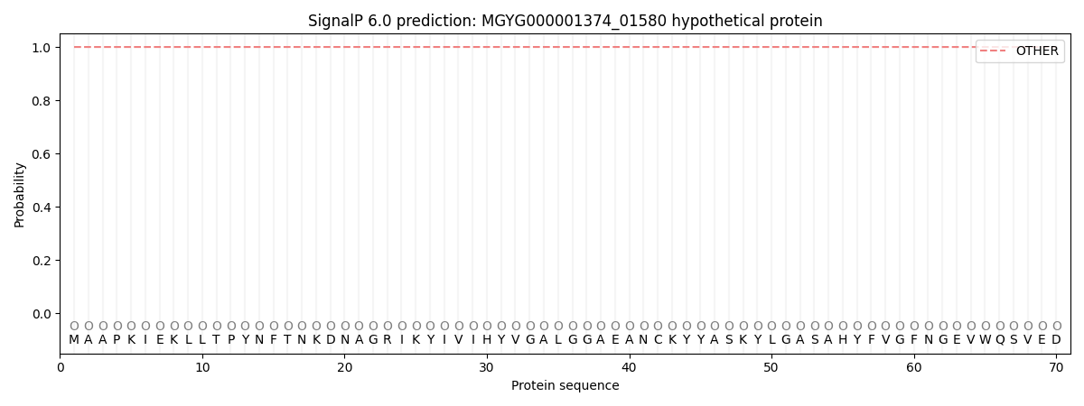
This protein is predicted as OTHER
| Other | SP_Sec_SPI | LIPO_Sec_SPII | TAT_Tat_SPI | TATLIP_Sec_SPII | PILIN_Sec_SPIII |
|---|---|---|---|---|---|
| 1.000094 | 0.000000 | 0.000000 | 0.000000 | 0.000000 | 0.000000 |