You are browsing environment: HUMAN GUT
CAZyme Information: MGYG000001451_04287
You are here: Home > Sequence: MGYG000001451_04287
Basic Information |
Genomic context |
Full Sequence |
Enzyme annotations |
CAZy signature domains |
CDD domains |
CAZyme hits |
PDB hits |
Swiss-Prot hits |
SignalP and Lipop annotations |
TMHMM annotations
Basic Information help
| Species | Paenibacillus_A antibioticophila | |||||||||||
|---|---|---|---|---|---|---|---|---|---|---|---|---|
| Lineage | Bacteria; Firmicutes; Bacilli; Paenibacillales; Paenibacillaceae; Paenibacillus_A; Paenibacillus_A antibioticophila | |||||||||||
| CAZyme ID | MGYG000001451_04287 | |||||||||||
| CAZy Family | GT4 | |||||||||||
| CAZyme Description | Spore coat protein SA | |||||||||||
| CAZyme Property |
|
|||||||||||
| Genome Property |
|
|||||||||||
| Gene Location | Start: 124433; End: 125584 Strand: - | |||||||||||
CDD Domains download full data without filtering help
| Cdd ID | Domain | E-Value | qStart | qEnd | sStart | sEnd | Domain Description |
|---|---|---|---|---|---|---|---|
| cd03801 | GT4_PimA-like | 5.18e-63 | 5 | 378 | 1 | 365 | phosphatidyl-myo-inositol mannosyltransferase. This family is most closely related to the GT4 family of glycosyltransferases and named after PimA in Propionibacterium freudenreichii, which is involved in the biosynthesis of phosphatidyl-myo-inositol mannosides (PIM) which are early precursors in the biosynthesis of lipomannans (LM) and lipoarabinomannans (LAM), and catalyzes the addition of a mannosyl residue from GDP-D-mannose (GDP-Man) to the position 2 of the carrier lipid phosphatidyl-myo-inositol (PI) to generate a phosphatidyl-myo-inositol bearing an alpha-1,2-linked mannose residue (PIM1). Glycosyltransferases catalyze the transfer of sugar moieties from activated donor molecules to specific acceptor molecules, forming glycosidic bonds. The acceptor molecule can be a lipid, a protein, a heterocyclic compound, or another carbohydrate residue. This group of glycosyltransferases is most closely related to the previously defined glycosyltransferase family 1 (GT1). The members of this family may transfer UDP, ADP, GDP, or CMP linked sugars. The diverse enzymatic activities among members of this family reflect a wide range of biological functions. The protein structure available for this family has the GTB topology, one of the two protein topologies observed for nucleotide-sugar-dependent glycosyltransferases. GTB proteins have distinct N- and C- terminal domains each containing a typical Rossmann fold. The two domains have high structural homology despite minimal sequence homology. The large cleft that separates the two domains includes the catalytic center and permits a high degree of flexibility. The members of this family are found mainly in certain bacteria and archaea. |
| pfam00534 | Glycos_transf_1 | 1.85e-45 | 195 | 359 | 1 | 157 | Glycosyl transferases group 1. Mutations in this domain of PIGA lead to disease (Paroxysmal Nocturnal haemoglobinuria). Members of this family transfer activated sugars to a variety of substrates, including glycogen, Fructose-6-phosphate and lipopolysaccharides. Members of this family transfer UDP, ADP, GDP or CMP linked sugars. The eukaryotic glycogen synthases may be distant members of this family. |
| COG0438 | RfaB | 6.84e-43 | 5 | 380 | 2 | 376 | Glycosyltransferase involved in cell wall bisynthesis [Cell wall/membrane/envelope biogenesis]. |
| cd03800 | GT4_sucrose_synthase | 1.22e-38 | 184 | 373 | 207 | 395 | sucrose-phosphate synthase and similar proteins. This family is most closely related to the GT4 family of glycosyltransferases. The sucrose-phosphate synthases in this family may be unique to plants and photosynthetic bacteria. This enzyme catalyzes the synthesis of sucrose 6-phosphate from fructose 6-phosphate and uridine 5'-diphosphate-glucose, a key regulatory step of sucrose metabolism. The activity of this enzyme is regulated by phosphorylation and moderated by the concentration of various metabolites and light. |
| pfam13692 | Glyco_trans_1_4 | 1.95e-36 | 196 | 346 | 1 | 138 | Glycosyl transferases group 1. |
CAZyme Hits help
| Hit ID | E-Value | Query Start | Query End | Hit Start | Hit End |
|---|---|---|---|---|---|
| ANS77253.1 | 8.06e-160 | 1 | 379 | 11 | 389 |
| AWB45820.1 | 7.72e-154 | 1 | 377 | 1 | 377 |
| AZS18439.1 | 9.09e-149 | 1 | 374 | 1 | 374 |
| ANY72476.1 | 3.85e-145 | 1 | 377 | 1 | 377 |
| AYB45887.1 | 5.11e-143 | 1 | 380 | 1 | 380 |
PDB Hits download full data without filtering help
| Hit ID | E-Value | Query Start | Query End | Hit Start | Hit End | Description |
|---|---|---|---|---|---|---|
| 6TVP_A | 9.45e-22 | 132 | 383 | 151 | 401 | Structureof Mycobacterium smegmatis alpha-maltose-1-phosphate synthase GlgM [Mycolicibacterium smegmatis MC2 155],6TVP_B Structure of Mycobacterium smegmatis alpha-maltose-1-phosphate synthase GlgM [Mycolicibacterium smegmatis MC2 155] |
| 3C4Q_A | 2.01e-19 | 136 | 379 | 166 | 403 | Structureof the retaining glycosyltransferase MshA : The first step in mycothiol biosynthesis. Organism : Corynebacterium glutamicum- Complex with UDP [Corynebacterium glutamicum],3C4Q_B Structure of the retaining glycosyltransferase MshA : The first step in mycothiol biosynthesis. Organism : Corynebacterium glutamicum- Complex with UDP [Corynebacterium glutamicum],3C4V_A Structure of the retaining glycosyltransferase MshA:The first step in mycothiol biosynthesis. Organism: Corynebacterium glutamicum : Complex with UDP and 1L-INS-1-P. [Corynebacterium glutamicum],3C4V_B Structure of the retaining glycosyltransferase MshA:The first step in mycothiol biosynthesis. Organism: Corynebacterium glutamicum : Complex with UDP and 1L-INS-1-P. [Corynebacterium glutamicum] |
| 3C48_A | 2.16e-19 | 136 | 379 | 186 | 423 | Structureof the retaining glycosyltransferase MshA: The first step in mycothiol biosynthesis. Organism: Corynebacterium glutamicum- APO (OPEN) structure. [Corynebacterium glutamicum],3C48_B Structure of the retaining glycosyltransferase MshA: The first step in mycothiol biosynthesis. Organism: Corynebacterium glutamicum- APO (OPEN) structure. [Corynebacterium glutamicum] |
| 3OKA_A | 2.44e-17 | 107 | 381 | 112 | 379 | Crystalstructure of Corynebacterium glutamicum PimB' in complex with GDP-Man (triclinic crystal form) [Corynebacterium glutamicum],3OKA_B Crystal structure of Corynebacterium glutamicum PimB' in complex with GDP-Man (triclinic crystal form) [Corynebacterium glutamicum] |
| 3OKC_A | 2.67e-17 | 107 | 381 | 112 | 379 | Crystalstructure of Corynebacterium glutamicum PimB' bound to GDP (orthorhombic crystal form) [Corynebacterium glutamicum],3OKP_A Crystal structure of Corynebacterium glutamicum PimB' bound to GDP-Man (orthorhombic crystal form) [Corynebacterium glutamicum] |
Swiss-Prot Hits download full data without filtering help
| Hit ID | E-Value | Query Start | Query End | Hit Start | Hit End | Description |
|---|---|---|---|---|---|---|
| P46915 | 9.35e-59 | 5 | 382 | 2 | 377 | Spore coat protein SA OS=Bacillus subtilis (strain 168) OX=224308 GN=cotSA PE=1 SV=1 |
| O34413 | 2.14e-52 | 5 | 380 | 2 | 375 | Putative glycosyltransferase YtcC OS=Bacillus subtilis (strain 168) OX=224308 GN=ytcC PE=3 SV=1 |
| D5UJ42 | 5.45e-23 | 136 | 379 | 201 | 437 | D-inositol 3-phosphate glycosyltransferase OS=Cellulomonas flavigena (strain ATCC 482 / DSM 20109 / BCRC 11376 / JCM 18109 / NBRC 3775 / NCIMB 8073 / NRS 134) OX=446466 GN=mshA PE=3 SV=1 |
| P26470 | 3.12e-21 | 74 | 374 | 90 | 371 | Lipopolysaccharide 1,2-N-acetylglucosaminetransferase OS=Salmonella typhimurium (strain LT2 / SGSC1412 / ATCC 700720) OX=99287 GN=waaK PE=3 SV=1 |
| D1BD84 | 3.31e-21 | 136 | 378 | 172 | 414 | D-inositol 3-phosphate glycosyltransferase OS=Sanguibacter keddieii (strain ATCC 51767 / DSM 10542 / NCFB 3025 / ST-74) OX=446469 GN=mshA PE=3 SV=1 |
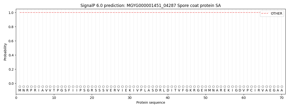
SignalP and Lipop Annotations help
This protein is predicted as OTHER
| Other | SP_Sec_SPI | LIPO_Sec_SPII | TAT_Tat_SPI | TATLIP_Sec_SPII | PILIN_Sec_SPIII |
|---|---|---|---|---|---|
| 1.000057 | 0.000000 | 0.000000 | 0.000000 | 0.000000 | 0.000000 |