You are browsing environment: HUMAN GUT
CAZyme Information: MGYG000001475_02462
You are here: Home > Sequence: MGYG000001475_02462
Basic Information |
Genomic context |
Full Sequence |
Enzyme annotations |
CAZy signature domains |
CDD domains |
CAZyme hits |
PDB hits |
Swiss-Prot hits |
SignalP and Lipop annotations |
TMHMM annotations
Basic Information help
| Species | Neobacillus rubiinfantis | |||||||||||
|---|---|---|---|---|---|---|---|---|---|---|---|---|
| Lineage | Bacteria; Firmicutes; Bacilli; Bacillales_B; DSM-18226; Neobacillus; Neobacillus rubiinfantis | |||||||||||
| CAZyme ID | MGYG000001475_02462 | |||||||||||
| CAZy Family | GT4 | |||||||||||
| CAZyme Description | Spore coat protein SA | |||||||||||
| CAZyme Property |
|
|||||||||||
| Genome Property |
|
|||||||||||
| Gene Location | Start: 58000; End: 59139 Strand: - | |||||||||||
CDD Domains download full data without filtering help
| Cdd ID | Domain | E-Value | qStart | qEnd | sStart | sEnd | Domain Description |
|---|---|---|---|---|---|---|---|
| cd03801 | GT4_PimA-like | 1.69e-68 | 2 | 376 | 1 | 365 | phosphatidyl-myo-inositol mannosyltransferase. This family is most closely related to the GT4 family of glycosyltransferases and named after PimA in Propionibacterium freudenreichii, which is involved in the biosynthesis of phosphatidyl-myo-inositol mannosides (PIM) which are early precursors in the biosynthesis of lipomannans (LM) and lipoarabinomannans (LAM), and catalyzes the addition of a mannosyl residue from GDP-D-mannose (GDP-Man) to the position 2 of the carrier lipid phosphatidyl-myo-inositol (PI) to generate a phosphatidyl-myo-inositol bearing an alpha-1,2-linked mannose residue (PIM1). Glycosyltransferases catalyze the transfer of sugar moieties from activated donor molecules to specific acceptor molecules, forming glycosidic bonds. The acceptor molecule can be a lipid, a protein, a heterocyclic compound, or another carbohydrate residue. This group of glycosyltransferases is most closely related to the previously defined glycosyltransferase family 1 (GT1). The members of this family may transfer UDP, ADP, GDP, or CMP linked sugars. The diverse enzymatic activities among members of this family reflect a wide range of biological functions. The protein structure available for this family has the GTB topology, one of the two protein topologies observed for nucleotide-sugar-dependent glycosyltransferases. GTB proteins have distinct N- and C- terminal domains each containing a typical Rossmann fold. The two domains have high structural homology despite minimal sequence homology. The large cleft that separates the two domains includes the catalytic center and permits a high degree of flexibility. The members of this family are found mainly in certain bacteria and archaea. |
| COG0438 | RfaB | 3.89e-43 | 1 | 379 | 1 | 377 | Glycosyltransferase involved in cell wall bisynthesis [Cell wall/membrane/envelope biogenesis]. |
| pfam00534 | Glycos_transf_1 | 5.39e-41 | 193 | 357 | 1 | 157 | Glycosyl transferases group 1. Mutations in this domain of PIGA lead to disease (Paroxysmal Nocturnal haemoglobinuria). Members of this family transfer activated sugars to a variety of substrates, including glycogen, Fructose-6-phosphate and lipopolysaccharides. Members of this family transfer UDP, ADP, GDP or CMP linked sugars. The eukaryotic glycogen synthases may be distant members of this family. |
| pfam13692 | Glyco_trans_1_4 | 1.25e-38 | 196 | 344 | 3 | 138 | Glycosyl transferases group 1. |
| cd03800 | GT4_sucrose_synthase | 5.26e-38 | 153 | 373 | 184 | 397 | sucrose-phosphate synthase and similar proteins. This family is most closely related to the GT4 family of glycosyltransferases. The sucrose-phosphate synthases in this family may be unique to plants and photosynthetic bacteria. This enzyme catalyzes the synthesis of sucrose 6-phosphate from fructose 6-phosphate and uridine 5'-diphosphate-glucose, a key regulatory step of sucrose metabolism. The activity of this enzyme is regulated by phosphorylation and moderated by the concentration of various metabolites and light. |
CAZyme Hits help
| Hit ID | E-Value | Query Start | Query End | Hit Start | Hit End |
|---|---|---|---|---|---|
| ASS95281.1 | 7.40e-210 | 1 | 378 | 1 | 378 |
| ALF10762.1 | 2.14e-209 | 1 | 379 | 1 | 379 |
| APM81577.1 | 2.14e-209 | 1 | 379 | 1 | 379 |
| ANZ30840.1 | 2.14e-209 | 1 | 379 | 1 | 379 |
| QYF83942.1 | 1.22e-208 | 1 | 378 | 1 | 378 |
PDB Hits download full data without filtering help
| Hit ID | E-Value | Query Start | Query End | Hit Start | Hit End | Description |
|---|---|---|---|---|---|---|
| 6TVP_A | 4.45e-18 | 129 | 378 | 150 | 398 | Structureof Mycobacterium smegmatis alpha-maltose-1-phosphate synthase GlgM [Mycolicibacterium smegmatis MC2 155],6TVP_B Structure of Mycobacterium smegmatis alpha-maltose-1-phosphate synthase GlgM [Mycolicibacterium smegmatis MC2 155] |
| 3OKA_A | 9.58e-14 | 128 | 356 | 132 | 356 | Crystalstructure of Corynebacterium glutamicum PimB' in complex with GDP-Man (triclinic crystal form) [Corynebacterium glutamicum],3OKA_B Crystal structure of Corynebacterium glutamicum PimB' in complex with GDP-Man (triclinic crystal form) [Corynebacterium glutamicum] |
| 3OKC_A | 1.02e-13 | 128 | 356 | 132 | 356 | Crystalstructure of Corynebacterium glutamicum PimB' bound to GDP (orthorhombic crystal form) [Corynebacterium glutamicum],3OKP_A Crystal structure of Corynebacterium glutamicum PimB' bound to GDP-Man (orthorhombic crystal form) [Corynebacterium glutamicum] |
| 3C4Q_A | 8.92e-12 | 157 | 379 | 189 | 405 | Structureof the retaining glycosyltransferase MshA : The first step in mycothiol biosynthesis. Organism : Corynebacterium glutamicum- Complex with UDP [Corynebacterium glutamicum],3C4Q_B Structure of the retaining glycosyltransferase MshA : The first step in mycothiol biosynthesis. Organism : Corynebacterium glutamicum- Complex with UDP [Corynebacterium glutamicum],3C4V_A Structure of the retaining glycosyltransferase MshA:The first step in mycothiol biosynthesis. Organism: Corynebacterium glutamicum : Complex with UDP and 1L-INS-1-P. [Corynebacterium glutamicum],3C4V_B Structure of the retaining glycosyltransferase MshA:The first step in mycothiol biosynthesis. Organism: Corynebacterium glutamicum : Complex with UDP and 1L-INS-1-P. [Corynebacterium glutamicum] |
| 3C48_A | 9.22e-12 | 157 | 379 | 209 | 425 | Structureof the retaining glycosyltransferase MshA: The first step in mycothiol biosynthesis. Organism: Corynebacterium glutamicum- APO (OPEN) structure. [Corynebacterium glutamicum],3C48_B Structure of the retaining glycosyltransferase MshA: The first step in mycothiol biosynthesis. Organism: Corynebacterium glutamicum- APO (OPEN) structure. [Corynebacterium glutamicum] |
Swiss-Prot Hits download full data without filtering help
| Hit ID | E-Value | Query Start | Query End | Hit Start | Hit End | Description |
|---|---|---|---|---|---|---|
| P46915 | 6.66e-107 | 1 | 378 | 1 | 375 | Spore coat protein SA OS=Bacillus subtilis (strain 168) OX=224308 GN=cotSA PE=1 SV=1 |
| O34413 | 1.52e-96 | 1 | 377 | 1 | 374 | Putative glycosyltransferase YtcC OS=Bacillus subtilis (strain 168) OX=224308 GN=ytcC PE=3 SV=1 |
| Q59002 | 2.01e-25 | 1 | 379 | 1 | 384 | Uncharacterized glycosyltransferase MJ1607 OS=Methanocaldococcus jannaschii (strain ATCC 43067 / DSM 2661 / JAL-1 / JCM 10045 / NBRC 100440) OX=243232 GN=MJ1607 PE=3 SV=1 |
| P26470 | 2.24e-24 | 2 | 369 | 4 | 368 | Lipopolysaccharide 1,2-N-acetylglucosaminetransferase OS=Salmonella typhimurium (strain LT2 / SGSC1412 / ATCC 700720) OX=99287 GN=waaK PE=3 SV=1 |
| A0R043 | 7.17e-23 | 14 | 375 | 9 | 372 | GDP-mannose-dependent alpha-(1-6)-phosphatidylinositol monomannoside mannosyltransferase OS=Mycolicibacterium smegmatis (strain ATCC 700084 / mc(2)155) OX=246196 GN=pimB PE=1 SV=1 |
SignalP and Lipop Annotations help
This protein is predicted as OTHER
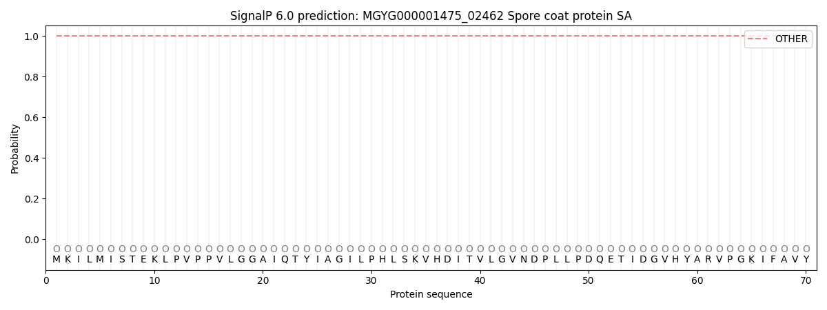
| Other | SP_Sec_SPI | LIPO_Sec_SPII | TAT_Tat_SPI | TATLIP_Sec_SPII | PILIN_Sec_SPIII |
|---|---|---|---|---|---|
| 1.000055 | 0.000000 | 0.000000 | 0.000000 | 0.000000 | 0.000000 |