You are browsing environment: HUMAN GUT
CAZyme Information: MGYG000001483_01191
You are here: Home > Sequence: MGYG000001483_01191
Basic Information |
Genomic context |
Full Sequence |
Enzyme annotations |
CAZy signature domains |
CDD domains |
CAZyme hits |
PDB hits |
Swiss-Prot hits |
SignalP and Lipop annotations |
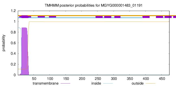
TMHMM annotations
Basic Information help
| Species | Arachnia massiliensis | |||||||||||
|---|---|---|---|---|---|---|---|---|---|---|---|---|
| Lineage | Bacteria; Actinobacteriota; Actinomycetia; Propionibacteriales; Propionibacteriaceae; Arachnia; Arachnia massiliensis | |||||||||||
| CAZyme ID | MGYG000001483_01191 | |||||||||||
| CAZy Family | GH109 | |||||||||||
| CAZyme Description | Glycosyl hydrolase family 109 protein 1 | |||||||||||
| CAZyme Property |
|
|||||||||||
| Genome Property |
|
|||||||||||
| Gene Location | Start: 247457; End: 248884 Strand: - | |||||||||||
CAZyme Signature Domains help
| Family | Start | End | Evalue | family coverage |
|---|---|---|---|---|
| GH109 | 65 | 459 | 7.2e-151 | 0.9874686716791979 |
CDD Domains download full data without filtering help
| Cdd ID | Domain | E-Value | qStart | qEnd | sStart | sEnd | Domain Description |
|---|---|---|---|---|---|---|---|
| COG0673 | MviM | 1.96e-25 | 67 | 259 | 3 | 182 | Predicted dehydrogenase [General function prediction only]. |
| pfam01408 | GFO_IDH_MocA | 7.11e-19 | 68 | 193 | 1 | 117 | Oxidoreductase family, NAD-binding Rossmann fold. This family of enzymes utilize NADP or NAD. This family is called the GFO/IDH/MOCA family in swiss-prot. |
| PRK11579 | PRK11579 | 3.09e-07 | 66 | 219 | 3 | 145 | putative oxidoreductase; Provisional |
| COG4091 | COG4091 | 1.20e-04 | 68 | 167 | 18 | 132 | Predicted homoserine dehydrogenase, contains C-terminal SAF domain [Amino acid transport and metabolism]. |
CAZyme Hits help
| Hit ID | E-Value | Query Start | Query End | Hit Start | Hit End |
|---|---|---|---|---|---|
| CQR65755.1 | 4.62e-231 | 1 | 473 | 1 | 471 |
| AJP00622.1 | 4.62e-231 | 1 | 473 | 1 | 471 |
| QNE79748.1 | 7.58e-231 | 8 | 475 | 8 | 477 |
| QIP83060.1 | 1.53e-230 | 8 | 475 | 8 | 477 |
| AYL34658.1 | 3.08e-230 | 8 | 475 | 8 | 477 |
PDB Hits download full data without filtering help
| Hit ID | E-Value | Query Start | Query End | Hit Start | Hit End | Description |
|---|---|---|---|---|---|---|
| 6T2B_A | 4.92e-99 | 49 | 459 | 24 | 439 | Glycosidehydrolase family 109 from Akkermansia muciniphila in complex with GalNAc and NAD+. [Akkermansia muciniphila],6T2B_B Glycoside hydrolase family 109 from Akkermansia muciniphila in complex with GalNAc and NAD+. [Akkermansia muciniphila],6T2B_C Glycoside hydrolase family 109 from Akkermansia muciniphila in complex with GalNAc and NAD+. [Akkermansia muciniphila],6T2B_D Glycoside hydrolase family 109 from Akkermansia muciniphila in complex with GalNAc and NAD+. [Akkermansia muciniphila] |
| 2IXA_A | 3.22e-71 | 62 | 459 | 11 | 432 | A-zyme,N-acetylgalactosaminidase [Elizabethkingia meningoseptica],2IXB_A Crystal structure of N-ACETYLGALACTOSAMINIDASE in complex with GalNAC [Elizabethkingia meningoseptica] |
| 3MOI_A | 9.25e-09 | 68 | 219 | 3 | 145 | Thecrystal structure of the putative dehydrogenase from Bordetella bronchiseptica RB50 [Bordetella bronchiseptica] |
| 6JW6_A | 2.87e-08 | 68 | 197 | 5 | 125 | Thecrystal structure of KanD2 in complex with NAD [Streptomyces kanamyceticus],6JW6_B The crystal structure of KanD2 in complex with NAD [Streptomyces kanamyceticus],6JW7_A The crystal structure of KanD2 in complex with NADH and 3'-deamino-3'-hydroxykanamycin A [Streptomyces kanamyceticus],6JW7_B The crystal structure of KanD2 in complex with NADH and 3'-deamino-3'-hydroxykanamycin A [Streptomyces kanamyceticus],6JW8_A The crystal structure of KanD2 in complex with NADH and 3'-deamino-3'-hydroxykanamycin B [Streptomyces kanamyceticus],6JW8_B The crystal structure of KanD2 in complex with NADH and 3'-deamino-3'-hydroxykanamycin B [Streptomyces kanamyceticus] |
| 3E82_A | 4.71e-08 | 66 | 219 | 6 | 148 | Crystalstructure of a putative oxidoreductase from Klebsiella pneumoniae [Micrococcus luteus NCTC 2665],3E82_B Crystal structure of a putative oxidoreductase from Klebsiella pneumoniae [Micrococcus luteus NCTC 2665],3E82_D Crystal structure of a putative oxidoreductase from Klebsiella pneumoniae [Micrococcus luteus NCTC 2665],3E82_E Crystal structure of a putative oxidoreductase from Klebsiella pneumoniae [Micrococcus luteus NCTC 2665] |
Swiss-Prot Hits download full data without filtering help
| Hit ID | E-Value | Query Start | Query End | Hit Start | Hit End | Description |
|---|---|---|---|---|---|---|
| Q9RK81 | 5.15e-226 | 1 | 473 | 1 | 469 | Glycosyl hydrolase family 109 protein OS=Streptomyces coelicolor (strain ATCC BAA-471 / A3(2) / M145) OX=100226 GN=SCO0529 PE=3 SV=1 |
| Q50HM6 | 7.21e-158 | 43 | 473 | 70 | 494 | Glycosyl hydrolase family 109 protein OS=Streptomyces niveus OX=193462 PE=3 SV=1 |
| B1W5J7 | 1.66e-156 | 49 | 467 | 77 | 489 | Glycosyl hydrolase family 109 protein OS=Streptomyces griseus subsp. griseus (strain JCM 4626 / NBRC 13350) OX=455632 GN=SGR_6325 PE=3 SV=1 |
| Q50EA3 | 9.45e-156 | 4 | 467 | 29 | 489 | Glycosyl hydrolase family 109 protein OS=Streptomyces filamentosus OX=67294 PE=3 SV=1 |
| A4FN60 | 6.22e-153 | 50 | 467 | 43 | 455 | Glycosyl hydrolase family 109 protein OS=Saccharopolyspora erythraea (strain ATCC 11635 / DSM 40517 / JCM 4748 / NBRC 13426 / NCIMB 8594 / NRRL 2338) OX=405948 GN=SACE_6314 PE=3 SV=1 |
SignalP and Lipop Annotations help
This protein is predicted as TAT

| Other | SP_Sec_SPI | LIPO_Sec_SPII | TAT_Tat_SPI | TATLIP_Sec_SPII | PILIN_Sec_SPIII |
|---|---|---|---|---|---|
| 0.000000 | 0.000000 | 0.000000 | 0.999951 | 0.000044 | 0.000000 |