You are browsing environment: HUMAN GUT
CAZyme Information: MGYG000001485_00476
You are here: Home > Sequence: MGYG000001485_00476
Basic Information |
Genomic context |
Full Sequence |
Enzyme annotations |
CAZy signature domains |
CDD domains |
CAZyme hits |
PDB hits |
Swiss-Prot hits |
SignalP and Lipop annotations |
TMHMM annotations
Basic Information help
| Species | Pseudomonas_E massiliensis | |||||||||||
|---|---|---|---|---|---|---|---|---|---|---|---|---|
| Lineage | Bacteria; Proteobacteria; Gammaproteobacteria; Pseudomonadales; Pseudomonadaceae; Pseudomonas_E; Pseudomonas_E massiliensis | |||||||||||
| CAZyme ID | MGYG000001485_00476 | |||||||||||
| CAZy Family | GT4 | |||||||||||
| CAZyme Description | Lipopolysaccharide core biosynthesis protein RfaG | |||||||||||
| CAZyme Property |
|
|||||||||||
| Genome Property |
|
|||||||||||
| Gene Location | Start: 216927; End: 218051 Strand: + | |||||||||||
CDD Domains download full data without filtering help
| Cdd ID | Domain | E-Value | qStart | qEnd | sStart | sEnd | Domain Description |
|---|---|---|---|---|---|---|---|
| cd03801 | GT4_PimA-like | 3.41e-42 | 2 | 368 | 1 | 362 | phosphatidyl-myo-inositol mannosyltransferase. This family is most closely related to the GT4 family of glycosyltransferases and named after PimA in Propionibacterium freudenreichii, which is involved in the biosynthesis of phosphatidyl-myo-inositol mannosides (PIM) which are early precursors in the biosynthesis of lipomannans (LM) and lipoarabinomannans (LAM), and catalyzes the addition of a mannosyl residue from GDP-D-mannose (GDP-Man) to the position 2 of the carrier lipid phosphatidyl-myo-inositol (PI) to generate a phosphatidyl-myo-inositol bearing an alpha-1,2-linked mannose residue (PIM1). Glycosyltransferases catalyze the transfer of sugar moieties from activated donor molecules to specific acceptor molecules, forming glycosidic bonds. The acceptor molecule can be a lipid, a protein, a heterocyclic compound, or another carbohydrate residue. This group of glycosyltransferases is most closely related to the previously defined glycosyltransferase family 1 (GT1). The members of this family may transfer UDP, ADP, GDP, or CMP linked sugars. The diverse enzymatic activities among members of this family reflect a wide range of biological functions. The protein structure available for this family has the GTB topology, one of the two protein topologies observed for nucleotide-sugar-dependent glycosyltransferases. GTB proteins have distinct N- and C- terminal domains each containing a typical Rossmann fold. The two domains have high structural homology despite minimal sequence homology. The large cleft that separates the two domains includes the catalytic center and permits a high degree of flexibility. The members of this family are found mainly in certain bacteria and archaea. |
| pfam00534 | Glycos_transf_1 | 2.57e-28 | 195 | 348 | 1 | 155 | Glycosyl transferases group 1. Mutations in this domain of PIGA lead to disease (Paroxysmal Nocturnal haemoglobinuria). Members of this family transfer activated sugars to a variety of substrates, including glycogen, Fructose-6-phosphate and lipopolysaccharides. Members of this family transfer UDP, ADP, GDP or CMP linked sugars. The eukaryotic glycogen synthases may be distant members of this family. |
| COG0438 | RfaB | 2.17e-21 | 1 | 368 | 1 | 371 | Glycosyltransferase involved in cell wall bisynthesis [Cell wall/membrane/envelope biogenesis]. |
| cd03800 | GT4_sucrose_synthase | 9.13e-21 | 154 | 367 | 181 | 396 | sucrose-phosphate synthase and similar proteins. This family is most closely related to the GT4 family of glycosyltransferases. The sucrose-phosphate synthases in this family may be unique to plants and photosynthetic bacteria. This enzyme catalyzes the synthesis of sucrose 6-phosphate from fructose 6-phosphate and uridine 5'-diphosphate-glucose, a key regulatory step of sucrose metabolism. The activity of this enzyme is regulated by phosphorylation and moderated by the concentration of various metabolites and light. |
| cd03821 | GT4_Bme6-like | 4.51e-16 | 114 | 368 | 140 | 376 | Brucella melitensis Bme6 and similar proteins. This family is most closely related to the GT4 family of glycosyltransferases. Bme6 in Brucella melitensis has been shown to be involved in the biosynthesis of a polysaccharide. |
CAZyme Hits help
| Hit ID | E-Value | Query Start | Query End | Hit Start | Hit End |
|---|---|---|---|---|---|
| ATR85100.1 | 1.90e-242 | 1 | 371 | 1 | 371 |
| VEE39021.1 | 3.15e-241 | 1 | 372 | 1 | 372 |
| QXZ03087.1 | 3.15e-241 | 1 | 372 | 1 | 372 |
| QOJ92203.1 | 3.15e-241 | 1 | 372 | 1 | 372 |
| AAN65974.1 | 3.15e-241 | 1 | 372 | 1 | 372 |
PDB Hits download full data without filtering help
| Hit ID | E-Value | Query Start | Query End | Hit Start | Hit End | Description |
|---|---|---|---|---|---|---|
| 2IW1_A | 2.02e-132 | 1 | 368 | 1 | 368 | CrystalStructure of WaaG, a glycosyltransferase involved in lipopolysaccharide biosynthesis [Escherichia coli str. K-12 substr. W3110] |
| 2IV7_A | 1.75e-128 | 3 | 368 | 3 | 368 | CrystalStructure of WaaG, a glycosyltransferase involved in lipopolysaccharide biosynthesis [Escherichia coli str. K-12 substr. W3110] |
| 2JJM_A | 4.37e-08 | 187 | 345 | 202 | 358 | CrystalStructure of a family GT4 glycosyltransferase from Bacillus anthracis ORF BA1558. [Bacillus anthracis str. Ames],2JJM_B Crystal Structure of a family GT4 glycosyltransferase from Bacillus anthracis ORF BA1558. [Bacillus anthracis str. Ames],2JJM_C Crystal Structure of a family GT4 glycosyltransferase from Bacillus anthracis ORF BA1558. [Bacillus anthracis str. Ames],2JJM_D Crystal Structure of a family GT4 glycosyltransferase from Bacillus anthracis ORF BA1558. [Bacillus anthracis str. Ames],2JJM_E Crystal Structure of a family GT4 glycosyltransferase from Bacillus anthracis ORF BA1558. [Bacillus anthracis str. Ames],2JJM_F Crystal Structure of a family GT4 glycosyltransferase from Bacillus anthracis ORF BA1558. [Bacillus anthracis str. Ames],2JJM_G Crystal Structure of a family GT4 glycosyltransferase from Bacillus anthracis ORF BA1558. [Bacillus anthracis str. Ames],2JJM_H Crystal Structure of a family GT4 glycosyltransferase from Bacillus anthracis ORF BA1558. [Bacillus anthracis str. Ames],2JJM_I Crystal Structure of a family GT4 glycosyltransferase from Bacillus anthracis ORF BA1558. [Bacillus anthracis str. Ames],2JJM_J Crystal Structure of a family GT4 glycosyltransferase from Bacillus anthracis ORF BA1558. [Bacillus anthracis str. Ames],2JJM_K Crystal Structure of a family GT4 glycosyltransferase from Bacillus anthracis ORF BA1558. [Bacillus anthracis str. Ames],2JJM_L Crystal Structure of a family GT4 glycosyltransferase from Bacillus anthracis ORF BA1558. [Bacillus anthracis str. Ames] |
| 3MBO_A | 4.58e-08 | 187 | 345 | 222 | 378 | CrystalStructure of the Glycosyltransferase BaBshA bound with UDP and L-malate [Bacillus anthracis],3MBO_B Crystal Structure of the Glycosyltransferase BaBshA bound with UDP and L-malate [Bacillus anthracis],3MBO_C Crystal Structure of the Glycosyltransferase BaBshA bound with UDP and L-malate [Bacillus anthracis],3MBO_D Crystal Structure of the Glycosyltransferase BaBshA bound with UDP and L-malate [Bacillus anthracis],3MBO_E Crystal Structure of the Glycosyltransferase BaBshA bound with UDP and L-malate [Bacillus anthracis],3MBO_F Crystal Structure of the Glycosyltransferase BaBshA bound with UDP and L-malate [Bacillus anthracis],3MBO_G Crystal Structure of the Glycosyltransferase BaBshA bound with UDP and L-malate [Bacillus anthracis],3MBO_H Crystal Structure of the Glycosyltransferase BaBshA bound with UDP and L-malate [Bacillus anthracis] |
| 4XSO_A | 2.34e-06 | 236 | 368 | 248 | 381 | ChainA, Alr3699 protein [Nostoc sp. PCC 7120 = FACHB-418],4XSO_B Chain B, Alr3699 protein [Nostoc sp. PCC 7120 = FACHB-418],4XSP_A Chain A, Alr3699 protein [Nostoc sp. PCC 7120 = FACHB-418],4XSP_B Chain B, Alr3699 protein [Nostoc sp. PCC 7120 = FACHB-418],4XSR_A Chain A, Alr3699 protein [Nostoc sp. PCC 7120 = FACHB-418],4XSR_B Chain B, Alr3699 protein [Nostoc sp. PCC 7120 = FACHB-418],4XSU_A Chain A, Alr3699 protein [Nostoc sp. PCC 7120 = FACHB-418],4XSU_B Chain B, Alr3699 protein [Nostoc sp. PCC 7120 = FACHB-418] |
Swiss-Prot Hits download full data without filtering help
| Hit ID | E-Value | Query Start | Query End | Hit Start | Hit End | Description |
|---|---|---|---|---|---|---|
| P25740 | 1.10e-131 | 1 | 368 | 1 | 368 | Lipopolysaccharide core biosynthesis protein RfaG OS=Escherichia coli (strain K12) OX=83333 GN=rfaG PE=1 SV=1 |
| B1MHQ0 | 1.53e-12 | 14 | 319 | 39 | 364 | D-inositol 3-phosphate glycosyltransferase OS=Mycobacteroides abscessus (strain ATCC 19977 / DSM 44196 / CIP 104536 / JCM 13569 / NCTC 13031 / TMC 1543) OX=561007 GN=mshA PE=3 SV=1 |
| Q5YP47 | 1.58e-11 | 14 | 352 | 34 | 397 | D-inositol 3-phosphate glycosyltransferase OS=Nocardia farcinica (strain IFM 10152) OX=247156 GN=mshA PE=3 SV=1 |
| D1BZ82 | 2.57e-11 | 152 | 342 | 187 | 381 | D-inositol 3-phosphate glycosyltransferase OS=Xylanimonas cellulosilytica (strain DSM 15894 / CECT 5975 / LMG 20990 / XIL07) OX=446471 GN=mshA PE=3 SV=1 |
| C8XA09 | 9.16e-10 | 14 | 318 | 49 | 379 | D-inositol 3-phosphate glycosyltransferase OS=Nakamurella multipartita (strain ATCC 700099 / DSM 44233 / CIP 104796 / JCM 9543 / NBRC 105858 / Y-104) OX=479431 GN=mshA PE=3 SV=1 |
SignalP and Lipop Annotations help
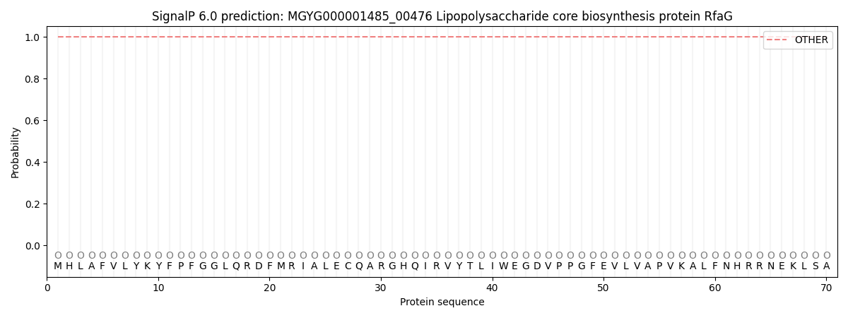
This protein is predicted as OTHER
| Other | SP_Sec_SPI | LIPO_Sec_SPII | TAT_Tat_SPI | TATLIP_Sec_SPII | PILIN_Sec_SPIII |
|---|---|---|---|---|---|
| 1.000067 | 0.000000 | 0.000000 | 0.000000 | 0.000000 | 0.000000 |