You are browsing environment: HUMAN GUT
CAZyme Information: MGYG000001486_01172
You are here: Home > Sequence: MGYG000001486_01172
Basic Information |
Genomic context |
Full Sequence |
Enzyme annotations |
CAZy signature domains |
CDD domains |
CAZyme hits |
PDB hits |
Swiss-Prot hits |
SignalP and Lipop annotations |
TMHMM annotations
Basic Information help
| Species | Mobilicoccus massiliensis | |||||||||||
|---|---|---|---|---|---|---|---|---|---|---|---|---|
| Lineage | Bacteria; Actinobacteriota; Actinomycetia; Actinomycetales; Dermatophilaceae; Mobilicoccus; Mobilicoccus massiliensis | |||||||||||
| CAZyme ID | MGYG000001486_01172 | |||||||||||
| CAZy Family | GT2 | |||||||||||
| CAZyme Description | Histidine biosynthesis bifunctional protein HisB | |||||||||||
| CAZyme Property |
|
|||||||||||
| Genome Property |
|
|||||||||||
| Gene Location | Start: 294579; End: 296315 Strand: + | |||||||||||
CAZyme Signature Domains help
| Family | Start | End | Evalue | family coverage |
|---|---|---|---|---|
| GT2 | 26 | 139 | 7.8e-17 | 0.6705882352941176 |
CDD Domains download full data without filtering help
| Cdd ID | Domain | E-Value | qStart | qEnd | sStart | sEnd | Domain Description |
|---|---|---|---|---|---|---|---|
| cd07503 | HAD_HisB-N | 6.35e-44 | 374 | 511 | 1 | 142 | histidinol phosphate phosphatase and related phosphatases. This family includes the N-terminal domain of the Escherichia coli bifunctional enzyme histidinol-phosphate phosphatase/imidazole-glycerol-phosphate dehydratase, HisB. The N-terminal histidinol-phosphate phosphatase domain catalyzes the dephosphorylation of histidinol phosphate, the eight step of L-histidine biosynthesis. This family also includes Escherichia coli GmhB phosphatase which is highly specific for D-glycero-D-manno-heptose-1,7-bisphosphate, it removes the C(7)phosphate and not the C(1)phosphate, and this is the third essential step of lipopolysaccharide heptose biosynthesis. This family belongs to the haloacid dehalogenase-like (HAD) hydrolases, a large superfamily of diverse enzymes that catalyze carbon or phosphoryl group transfer reactions on a range of substrates, using an active site aspartate in nucleophilic catalysis. Members of this superfamily include 2-L-haloalkanoic acid dehalogenase, azetidine hydrolase, phosphonoacetaldehyde hydrolase, phosphoserine phosphatase, phosphomannomutase, P-type ATPases and many others. HAD hydrolases are found in all three kingdoms of life, and most genomes are predicted to contain multiple HAD-like proteins. Members possess a highly conserved alpha/beta core domain, and many also possess a small cap domain, the fold and function of which is variable. HAD hydrolases are sometimes referred to as belonging to the DDDD superfamily of phosphohydrolases. |
| PRK08942 | PRK08942 | 2.49e-29 | 371 | 543 | 1 | 178 | D-glycero-beta-D-manno-heptose 1,7-bisphosphate 7-phosphatase. |
| COG0241 | HisB1 | 2.35e-28 | 373 | 526 | 5 | 162 | Histidinol phosphatase or a related phosphatase [Amino acid transport and metabolism]. |
| TIGR01656 | Histidinol-ppas | 2.59e-26 | 375 | 513 | 2 | 146 | histidinol-phosphate phosphatase family domain. This domain is found in authentic histidinol-phosphate phosphatases which are sometimes found as stand-alone entities and sometimes as fusions with imidazoleglycerol-phosphate dehydratase (TIGR01261). Additionally, a family of proteins including YaeD from E. coli (TIGR00213) and various other proteins are closely related but may not have the same substrate specificity. This domain is a member of the haloacid-dehalogenase (HAD) superfamily of aspartate-nucleophile hydrolases. This superfamily is distinguished by the presence of three motifs: an N-terminal motif containing the nucleophilic aspartate, a central motif containing an conserved serine or threonine, and a C-terminal motif containing a conserved lysine (or arginine) and conserved aspartates. More specifically, the domian modelled here is a member of subfamily III of the HAD-superfamily by virtue of lacking a "capping" domain in either of the two common positions, between motifs 1 and 2, or between motifs 2 and 3. |
| TIGR01662 | HAD-SF-IIIA | 2.26e-25 | 374 | 513 | 1 | 135 | HAD-superfamily hydrolase, subfamily IIIA. This subfamily falls within the Haloacid Dehalogenase (HAD) superfamily of aspartate-nucleophile hydrolases. The Class III subfamilies are characterized by the lack of any domains located between either between the first and second conserved catalytic motifs (as in the Class I subfamilies, TIGR01493, TIGR01509, TIGR01488 and TIGR01494) or between the second and third conserved catalytic motifs (as in the Class II subfamilies, TIGR01460 and TIGR01484) of the superfamily domain. The IIIA subfamily contains five major clades: histidinol-phosphatase (TIGR01261) and histidinol-phosphatase-related protein (TIGR00213) which together form a subfamily (TIGR01656), DNA 3'-phosphatase (TIGR01663, TIGR01664), YqeG (TIGR01668) and YrbI (TIGR01670). In the case of histidinol phosphatase and PNK-3'-phosphatase, this model represents a domain of a bifunctional system. In the histidinol phosphatase HisB, a C-terminal domain is an imidazoleglycerol-phosphate dehydratase which catalyzes a related step in histidine biosynthesis. In PNK-3'-phosphatase, N- and C-terminal domains constitute the polynucleotide kinase and DNA-binding components of the enzyme. [Unknown function, Enzymes of unknown specificity] |
CAZyme Hits help
| Hit ID | E-Value | Query Start | Query End | Hit Start | Hit End |
|---|---|---|---|---|---|
| AVM63328.1 | 1.17e-186 | 24 | 535 | 6 | 536 |
| QCU78081.1 | 4.90e-186 | 25 | 541 | 15 | 527 |
| QDP77205.1 | 8.80e-186 | 25 | 535 | 3 | 506 |
| ARK05330.1 | 3.55e-185 | 25 | 535 | 3 | 506 |
| QCB93721.1 | 5.44e-181 | 17 | 539 | 4 | 511 |
PDB Hits download full data without filtering help
| Hit ID | E-Value | Query Start | Query End | Hit Start | Hit End | Description |
|---|---|---|---|---|---|---|
| 4PNH_A | 9.69e-13 | 368 | 517 | 14 | 168 | Crystalstructure of D,D-heptose 1,7-bisphosphate phosphatase from Burkholderia Thailandensis [Burkholderia thailandensis E264],4PNH_B Crystal structure of D,D-heptose 1,7-bisphosphate phosphatase from Burkholderia Thailandensis [Burkholderia thailandensis E264],4PNH_C Crystal structure of D,D-heptose 1,7-bisphosphate phosphatase from Burkholderia Thailandensis [Burkholderia thailandensis E264],4PNH_D Crystal structure of D,D-heptose 1,7-bisphosphate phosphatase from Burkholderia Thailandensis [Burkholderia thailandensis E264],4PNH_E Crystal structure of D,D-heptose 1,7-bisphosphate phosphatase from Burkholderia Thailandensis [Burkholderia thailandensis E264],4PNH_F Crystal structure of D,D-heptose 1,7-bisphosphate phosphatase from Burkholderia Thailandensis [Burkholderia thailandensis E264],4PNH_G Crystal structure of D,D-heptose 1,7-bisphosphate phosphatase from Burkholderia Thailandensis [Burkholderia thailandensis E264],4PNH_H Crystal structure of D,D-heptose 1,7-bisphosphate phosphatase from Burkholderia Thailandensis [Burkholderia thailandensis E264],4PNH_I Crystal structure of D,D-heptose 1,7-bisphosphate phosphatase from Burkholderia Thailandensis [Burkholderia thailandensis E264],4PNH_J Crystal structure of D,D-heptose 1,7-bisphosphate phosphatase from Burkholderia Thailandensis [Burkholderia thailandensis E264],4PNH_K Crystal structure of D,D-heptose 1,7-bisphosphate phosphatase from Burkholderia Thailandensis [Burkholderia thailandensis E264],4PNH_L Crystal structure of D,D-heptose 1,7-bisphosphate phosphatase from Burkholderia Thailandensis [Burkholderia thailandensis E264] |
| 3L8E_A | 8.83e-12 | 375 | 533 | 7 | 178 | CrystalStructure of apo form of D,D-heptose 1.7-bisphosphate phosphatase from E. Coli [Escherichia coli K-12],3L8E_B Crystal Structure of apo form of D,D-heptose 1.7-bisphosphate phosphatase from E. Coli [Escherichia coli K-12] |
| 2GMW_A | 1.36e-11 | 375 | 533 | 27 | 198 | CrystalStructure of D,D-heptose 1.7-bisphosphate phosphatase from E. Coli. [Escherichia coli],2GMW_B Crystal Structure of D,D-heptose 1.7-bisphosphate phosphatase from E. Coli. [Escherichia coli],3ESQ_A Crystal Structure of Calcium-bound D,D-heptose 1.7-bisphosphate phosphatase from E. Coli [Escherichia coli K-12],3ESR_A Crystal Structure of D,D-heptose1.7-bisphosphate phosphatase from E. coli in complex with calcium and phosphate [Escherichia coli K-12],3L1U_A Crystal structure of Calcium-bound GmhB from E. coli. [Escherichia coli K-12],3L1U_B Crystal structure of Calcium-bound GmhB from E. coli. [Escherichia coli K-12],3L1V_A Crystal structure of GmhB from E. coli in complex with calcium and phosphate. [Escherichia coli K-12],3L1V_B Crystal structure of GmhB from E. coli in complex with calcium and phosphate. [Escherichia coli K-12] |
| 3L8H_A | 1.89e-11 | 374 | 517 | 2 | 150 | CrystalStructure of D,D-heptose 1.7-bisphosphate phosphatase from B. bronchiseptica complexed with magnesium and phosphate [Bordetella bronchiseptica],3L8H_B Crystal Structure of D,D-heptose 1.7-bisphosphate phosphatase from B. bronchiseptica complexed with magnesium and phosphate [Bordetella bronchiseptica],3L8H_C Crystal Structure of D,D-heptose 1.7-bisphosphate phosphatase from B. bronchiseptica complexed with magnesium and phosphate [Bordetella bronchiseptica],3L8H_D Crystal Structure of D,D-heptose 1.7-bisphosphate phosphatase from B. bronchiseptica complexed with magnesium and phosphate [Bordetella bronchiseptica] |
| 3L8F_A | 2.52e-10 | 375 | 533 | 7 | 178 | CrystalStructure of D,D-heptose 1.7-bisphosphate phosphatase from E. Coli complexed with magnesium and phosphate [Escherichia coli K-12],3L8G_A Crystal Structure of D,D-heptose 1.7-bisphosphate phosphatase from E. Coli complexed with D-glycero-D-manno-heptose 1, 7-bisphosphate [Escherichia coli K-12] |
Swiss-Prot Hits download full data without filtering help
| Hit ID | E-Value | Query Start | Query End | Hit Start | Hit End | Description |
|---|---|---|---|---|---|---|
| Q88RS0 | 8.19e-16 | 374 | 513 | 2 | 146 | D-glycero-beta-D-manno-heptose-1,7-bisphosphate 7-phosphatase OS=Pseudomonas putida (strain ATCC 47054 / DSM 6125 / CFBP 8728 / NCIMB 11950 / KT2440) OX=160488 GN=gmhB PE=1 SV=1 |
| Q9I7C0 | 5.55e-13 | 376 | 513 | 6 | 148 | D-glycero-beta-D-manno-heptose-1,7-bisphosphate 7-phosphatase OS=Pseudomonas aeruginosa (strain ATCC 15692 / DSM 22644 / CIP 104116 / JCM 14847 / LMG 12228 / 1C / PRS 101 / PAO1) OX=208964 GN=gmhB PE=3 SV=1 |
| Q9HKQ2 | 1.28e-12 | 374 | 503 | 16 | 148 | Probable D-glycero-D-manno-heptose-1,7-bisphosphate 7-phosphatase OS=Thermoplasma acidophilum (strain ATCC 25905 / DSM 1728 / JCM 9062 / NBRC 15155 / AMRC-C165) OX=273075 GN=gmhB PE=3 SV=1 |
| Q97B60 | 2.33e-12 | 374 | 493 | 2 | 125 | Probable D-glycero-D-manno-heptose-1,7-bisphosphate 7-phosphatase OS=Thermoplasma volcanium (strain ATCC 51530 / DSM 4299 / JCM 9571 / NBRC 15438 / GSS1) OX=273116 GN=gmhB PE=3 SV=1 |
| Q7VL21 | 9.51e-12 | 374 | 520 | 4 | 162 | D-glycero-beta-D-manno-heptose-1,7-bisphosphate 7-phosphatase OS=Haemophilus ducreyi (strain 35000HP / ATCC 700724) OX=233412 GN=gmhB PE=3 SV=1 |
SignalP and Lipop Annotations help
This protein is predicted as OTHER
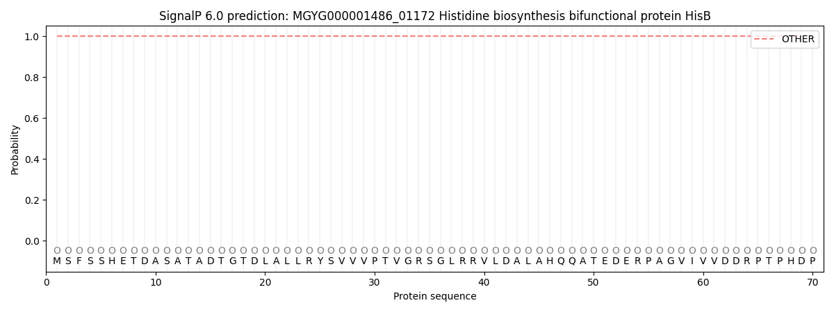
| Other | SP_Sec_SPI | LIPO_Sec_SPII | TAT_Tat_SPI | TATLIP_Sec_SPII | PILIN_Sec_SPIII |
|---|---|---|---|---|---|
| 1.000014 | 0.000012 | 0.000000 | 0.000000 | 0.000000 | 0.000000 |