You are browsing environment: HUMAN GUT
CAZyme Information: MGYG000001507_00994
You are here: Home > Sequence: MGYG000001507_00994
Basic Information |
Genomic context |
Full Sequence |
Enzyme annotations |
CAZy signature domains |
CDD domains |
CAZyme hits |
PDB hits |
Swiss-Prot hits |
SignalP and Lipop annotations |
TMHMM annotations
Basic Information help
| Species | Paenibacillus ihuae | |||||||||||
|---|---|---|---|---|---|---|---|---|---|---|---|---|
| Lineage | Bacteria; Firmicutes; Bacilli; Paenibacillales; Paenibacillaceae; Paenibacillus; Paenibacillus ihuae | |||||||||||
| CAZyme ID | MGYG000001507_00994 | |||||||||||
| CAZy Family | CBM22 | |||||||||||
| CAZyme Description | hypothetical protein | |||||||||||
| CAZyme Property |
|
|||||||||||
| Genome Property |
|
|||||||||||
| Gene Location | Start: 1070285; End: 1076443 Strand: + | |||||||||||
CAZyme Signature Domains help
| Family | Start | End | Evalue | family coverage |
|---|---|---|---|---|
| GH10 | 367 | 696 | 9e-100 | 0.9867986798679867 |
| GH10 | 1081 | 1411 | 2e-99 | 0.9900990099009901 |
| CBM9 | 723 | 895 | 8.6e-57 | 0.9945054945054945 |
| CBM9 | 1438 | 1610 | 5.4e-56 | 0.9945054945054945 |
| CBM22 | 206 | 334 | 1.5e-31 | 0.9465648854961832 |
CDD Domains download full data without filtering help
| Cdd ID | Domain | E-Value | qStart | qEnd | sStart | sEnd | Domain Description |
|---|---|---|---|---|---|---|---|
| pfam00331 | Glyco_hydro_10 | 8.29e-111 | 1082 | 1411 | 2 | 310 | Glycosyl hydrolase family 10. |
| pfam00331 | Glyco_hydro_10 | 1.13e-110 | 367 | 696 | 2 | 310 | Glycosyl hydrolase family 10. |
| smart00633 | Glyco_10 | 2.56e-97 | 406 | 694 | 1 | 263 | Glycosyl hydrolase family 10. |
| smart00633 | Glyco_10 | 2.02e-91 | 1121 | 1409 | 1 | 263 | Glycosyl hydrolase family 10. |
| cd00005 | CBM9_like_1 | 4.46e-91 | 713 | 896 | 1 | 185 | DOMON-like type 9 carbohydrate binding module of xylanases. Family 9 carbohydrate-binding modules (CBM9) play a role in the microbial degradation of cellulose and hemicellulose (materials found in plants). The domain has previously been called cellulose-binding domain. The polysaccharide binding sites of CBMs with available 3D structure have been found to be either flat surfaces with interactions formed by predominantly aromatic residues (tryptophan and tyrosine), or extended shallow grooves. The CBM9 domain frequently occurs in tandem repeats; members found in this subfamily typically co-occur with glycosyl hydrolase family 10 domains and are annotated as endo-1,4-beta-xylanases. CBM9 from Thermotoga maritima xylanase 10A is reported to have specificity for polysaccharide reducing ends. |
CAZyme Hits help
| Hit ID | E-Value | Query Start | Query End | Hit Start | Hit End |
|---|---|---|---|---|---|
| QSF46386.1 | 0.0 | 1 | 2052 | 1 | 1339 |
| AIQ51609.1 | 0.0 | 1 | 2052 | 1 | 1340 |
| AIQ45999.1 | 0.0 | 1 | 2052 | 1 | 1340 |
| AIQ57217.1 | 0.0 | 1 | 2052 | 1 | 1343 |
| AIQ28411.1 | 0.0 | 1 | 2052 | 1 | 1346 |
PDB Hits download full data without filtering help
| Hit ID | E-Value | Query Start | Query End | Hit Start | Hit End | Description |
|---|---|---|---|---|---|---|
| 3RDK_A | 1.67e-179 | 363 | 699 | 4 | 340 | Proteincrystal structure of xylanase A1 of Paenibacillus sp. JDR-2 [Paenibacillus sp. JDR-2],3RDK_B Protein crystal structure of xylanase A1 of Paenibacillus sp. JDR-2 [Paenibacillus sp. JDR-2],3RO8_A Crystal structure of the catalytic domain of XynA1 from Paenibacillus sp. JDR-2 [Paenibacillus sp. JDR-2],3RO8_B Crystal structure of the catalytic domain of XynA1 from Paenibacillus sp. JDR-2 [Paenibacillus sp. JDR-2],3RO8_C Crystal structure of the catalytic domain of XynA1 from Paenibacillus sp. JDR-2 [Paenibacillus sp. JDR-2],3RO8_D Crystal structure of the catalytic domain of XynA1 from Paenibacillus sp. JDR-2 [Paenibacillus sp. JDR-2],3RO8_E Crystal structure of the catalytic domain of XynA1 from Paenibacillus sp. JDR-2 [Paenibacillus sp. JDR-2],3RO8_F Crystal structure of the catalytic domain of XynA1 from Paenibacillus sp. JDR-2 [Paenibacillus sp. JDR-2],3RO8_G Crystal structure of the catalytic domain of XynA1 from Paenibacillus sp. JDR-2 [Paenibacillus sp. JDR-2],3RO8_H Crystal structure of the catalytic domain of XynA1 from Paenibacillus sp. JDR-2 [Paenibacillus sp. JDR-2],4E4P_A Second native structure of Xylanase A1 from Paenibacillus sp. JDR-2 [Paenibacillus sp. JDR-2],4E4P_B Second native structure of Xylanase A1 from Paenibacillus sp. JDR-2 [Paenibacillus sp. JDR-2] |
| 3W24_A | 3.17e-72 | 1074 | 1411 | 2 | 325 | Thehigh-resolution crystal structure of TsXylA, intracellular xylanase from Thermoanaerobacterium saccharolyticum JW/SL-YS485 [Thermoanaerobacterium saccharolyticum JW/SL-YS485] |
| 3W27_A | 2.01e-71 | 1074 | 1411 | 2 | 325 | Thehigh-resolution crystal structure of TsXylA, intracellular xylanase from /Thermoanaerobacterium saccharolyticum JW/SL-YS485/: the complex of the E251A mutant with xylobiose [Thermoanaerobacterium saccharolyticum JW/SL-YS485],3W28_A The high-resolution crystal structure of TsXylA, intracellular xylanase from /Thermoanaerobacterium saccharolyticum JW/SL-YS485/: the complex of the E251A mutant with xylotriose [Thermoanaerobacterium saccharolyticum JW/SL-YS485],3W29_A The high-resolution crystal structure of TsXylA, intracellular xylanase from /Thermoanaerobacterium saccharolyticum JW/SL-YS485/: the complex of the E251A mutant with xylotetraose [Thermoanaerobacterium saccharolyticum JW/SL-YS485] |
| 3W25_A | 2.01e-71 | 1074 | 1411 | 2 | 325 | Thehigh-resolution crystal structure of TsXylA, intracellular xylanase from /Thermoanaerobacterium saccharolyticum JW/SL-YS485/: the complex of the E146A mutant with xylobiose [Thermoanaerobacterium saccharolyticum JW/SL-YS485],3W26_A The high-resolution crystal structure of TsXylA, intracellular xylanase from /Thermoanaerobacterium saccharolyticum JW/SL-YS485/: the complex of the E146A mutant with xylotriose [Thermoanaerobacterium saccharolyticum JW/SL-YS485] |
| 4HU8_A | 4.15e-71 | 1077 | 1410 | 95 | 441 | CrystalStructure of a Bacterial Ig-like Domain Containing GH10 Xylanase from Termite Gut [Globitermes brachycerastes],4HU8_B Crystal Structure of a Bacterial Ig-like Domain Containing GH10 Xylanase from Termite Gut [Globitermes brachycerastes],4HU8_C Crystal Structure of a Bacterial Ig-like Domain Containing GH10 Xylanase from Termite Gut [Globitermes brachycerastes],4HU8_D Crystal Structure of a Bacterial Ig-like Domain Containing GH10 Xylanase from Termite Gut [Globitermes brachycerastes],4HU8_E Crystal Structure of a Bacterial Ig-like Domain Containing GH10 Xylanase from Termite Gut [Globitermes brachycerastes],4HU8_F Crystal Structure of a Bacterial Ig-like Domain Containing GH10 Xylanase from Termite Gut [Globitermes brachycerastes],4HU8_G Crystal Structure of a Bacterial Ig-like Domain Containing GH10 Xylanase from Termite Gut [Globitermes brachycerastes],4HU8_H Crystal Structure of a Bacterial Ig-like Domain Containing GH10 Xylanase from Termite Gut [Globitermes brachycerastes] |
Swiss-Prot Hits download full data without filtering help
| Hit ID | E-Value | Query Start | Query End | Hit Start | Hit End | Description |
|---|---|---|---|---|---|---|
| C6CRV0 | 0.0 | 38 | 2052 | 189 | 1461 | Endo-1,4-beta-xylanase A OS=Paenibacillus sp. (strain JDR-2) OX=324057 GN=xynA1 PE=1 SV=1 |
| P38535 | 2.68e-123 | 208 | 900 | 45 | 899 | Exoglucanase XynX OS=Acetivibrio thermocellus OX=1515 GN=xynX PE=3 SV=1 |
| P36917 | 3.13e-121 | 922 | 1614 | 202 | 1046 | Endo-1,4-beta-xylanase A OS=Thermoanaerobacterium saccharolyticum OX=28896 GN=xynA PE=1 SV=1 |
| Q60042 | 5.22e-106 | 804 | 1610 | 77 | 1053 | Endo-1,4-beta-xylanase A OS=Thermotoga neapolitana OX=2337 GN=xynA PE=1 SV=1 |
| Q60037 | 2.73e-104 | 936 | 1610 | 222 | 1057 | Endo-1,4-beta-xylanase A OS=Thermotoga maritima (strain ATCC 43589 / DSM 3109 / JCM 10099 / NBRC 100826 / MSB8) OX=243274 GN=xynA PE=1 SV=1 |
SignalP and Lipop Annotations help
This protein is predicted as SP
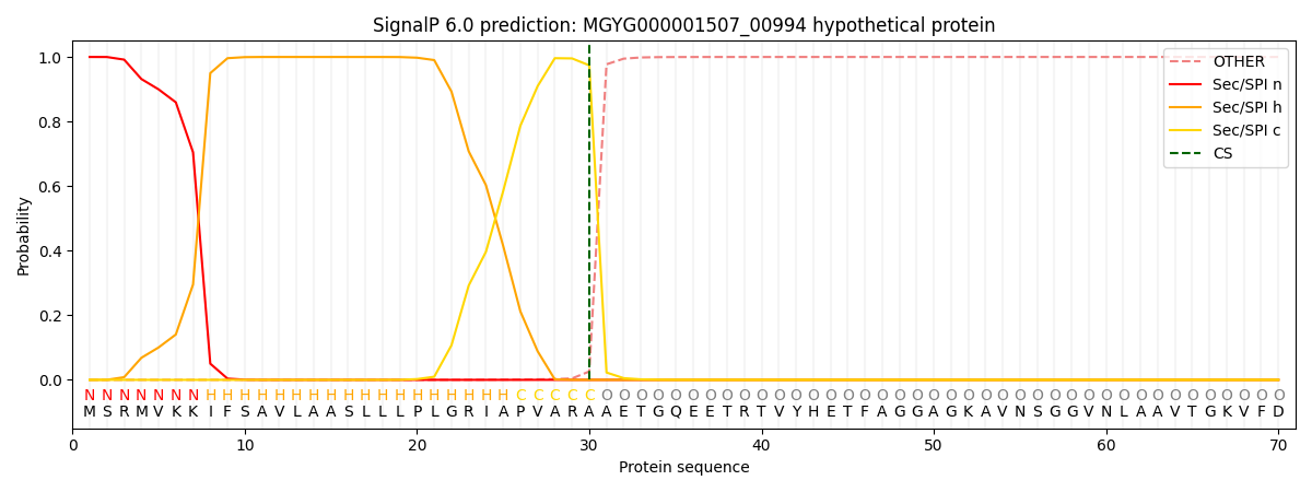
| Other | SP_Sec_SPI | LIPO_Sec_SPII | TAT_Tat_SPI | TATLIP_Sec_SPII | PILIN_Sec_SPIII |
|---|---|---|---|---|---|
| 0.000278 | 0.998974 | 0.000191 | 0.000186 | 0.000178 | 0.000161 |