You are browsing environment: HUMAN GUT
CAZyme Information: MGYG000001507_05887
You are here: Home > Sequence: MGYG000001507_05887
Basic Information |
Genomic context |
Full Sequence |
Enzyme annotations |
CAZy signature domains |
CDD domains |
CAZyme hits |
PDB hits |
Swiss-Prot hits |
SignalP and Lipop annotations |
TMHMM annotations
Basic Information help
| Species | Paenibacillus ihuae | |||||||||||
|---|---|---|---|---|---|---|---|---|---|---|---|---|
| Lineage | Bacteria; Firmicutes; Bacilli; Paenibacillales; Paenibacillaceae; Paenibacillus; Paenibacillus ihuae | |||||||||||
| CAZyme ID | MGYG000001507_05887 | |||||||||||
| CAZy Family | GT2 | |||||||||||
| CAZyme Description | hypothetical protein | |||||||||||
| CAZyme Property |
|
|||||||||||
| Genome Property |
|
|||||||||||
| Gene Location | Start: 99970; End: 101787 Strand: - | |||||||||||
CAZyme Signature Domains help
| Family | Start | End | Evalue | family coverage |
|---|---|---|---|---|
| GT2 | 207 | 322 | 3e-23 | 0.6764705882352942 |
CDD Domains download full data without filtering help
| Cdd ID | Domain | E-Value | qStart | qEnd | sStart | sEnd | Domain Description |
|---|---|---|---|---|---|---|---|
| pfam00535 | Glycos_transf_2 | 1.50e-22 | 207 | 321 | 1 | 114 | Glycosyl transferase family 2. Diverse family, transferring sugar from UDP-glucose, UDP-N-acetyl- galactosamine, GDP-mannose or CDP-abequose, to a range of substrates including cellulose, dolichol phosphate and teichoic acids. |
| cd00761 | Glyco_tranf_GTA_type | 1.08e-18 | 208 | 311 | 1 | 103 | Glycosyltransferase family A (GT-A) includes diverse families of glycosyl transferases with a common GT-A type structural fold. Glycosyltransferases (GTs) are enzymes that synthesize oligosaccharides, polysaccharides, and glycoconjugates by transferring the sugar moiety from an activated nucleotide-sugar donor to an acceptor molecule, which may be a growing oligosaccharide, a lipid, or a protein. Based on the stereochemistry of the donor and acceptor molecules, GTs are classified as either retaining or inverting enzymes. To date, all GT structures adopt one of two possible folds, termed GT-A fold and GT-B fold. This hierarchy includes diverse families of glycosyl transferases with a common GT-A type structural fold, which has two tightly associated beta/alpha/beta domains that tend to form a continuous central sheet of at least eight beta-strands. The majority of the proteins in this superfamily are Glycosyltransferase family 2 (GT-2) proteins. But it also includes families GT-43, GT-6, GT-8, GT13 and GT-7; which are evolutionarily related to GT-2 and share structure similarities. |
| cd02525 | Succinoglycan_BP_ExoA | 8.88e-17 | 206 | 482 | 2 | 223 | ExoA is involved in the biosynthesis of succinoglycan. Succinoglycan Biosynthesis Protein ExoA catalyzes the formation of a beta-1,3 linkage of the second sugar (glucose) of the succinoglycan with the galactose on the lipid carrie. Succinoglycan is an acidic exopolysaccharide that is important for invasion of the nodules. Succinoglycan is a high-molecular-weight polymer composed of repeating octasaccharide units. These units are synthesized on membrane-bound isoprenoid lipid carriers, beginning with galactose followed by seven glucose molecules, and modified by the addition of acetate, succinate, and pyruvate. ExoA is a membrane protein with a transmembrance domain at c-terminus. |
| cd06420 | GT2_Chondriotin_Pol_N | 7.27e-16 | 208 | 456 | 1 | 169 | N-terminal domain of Chondroitin polymerase functions as a GalNAc transferase. Chondroitin polymerase is a two domain, bi-functional protein. The N-terminal domain functions as a GalNAc transferase. The bacterial chondroitin polymerase catalyzes elongation of the chondroitin chain by alternatively transferring the GlcUA and GalNAc moiety from UDP-GlcUA and UDP-GalNAc to the non-reducing ends of the chondroitin chain. The enzyme consists of N-terminal and C-terminal domains in which the two active sites catalyze the addition of GalNAc and GlcUA, respectively. Chondroitin chains range from 40 to over 100 repeating units of the disaccharide. Sulfated chondroitins are involved in the regulation of various biological functions such as central nervous system development, wound repair, infection, growth factor signaling, and morphogenesis, in addition to its conventional structural roles. In Caenorhabditis elegans, chondroitin is an essential factor for the worm to undergo cytokinesis and cell division. Chondroitin is synthesized as proteoglycans, sulfated and secreted to the cell surface or extracellular matrix. |
| cd06423 | CESA_like | 2.57e-13 | 208 | 324 | 1 | 118 | CESA_like is the cellulose synthase superfamily. The cellulose synthase (CESA) superfamily includes a wide variety of glycosyltransferase family 2 enzymes that share the common characteristic of catalyzing the elongation of polysaccharide chains. The members include cellulose synthase catalytic subunit, chitin synthase, glucan biosynthesis protein and other families of CESA-like proteins. Cellulose synthase catalyzes the polymerization reaction of cellulose, an aggregate of unbranched polymers of beta-1,4-linked glucose residues in plants, most algae, some bacteria and fungi, and even some animals. In bacteria, algae and lower eukaryotes, there is a second unrelated type of cellulose synthase (Type II), which produces acylated cellulose, a derivative of cellulose. Chitin synthase catalyzes the incorporation of GlcNAc from substrate UDP-GlcNAc into chitin, which is a linear homopolymer of beta-(1,4)-linked GlcNAc residues and Glucan Biosynthesis protein catalyzes the elongation of beta-1,2 polyglucose chains of Glucan. |
CAZyme Hits help
| Hit ID | E-Value | Query Start | Query End | Hit Start | Hit End |
|---|---|---|---|---|---|
| QWU15230.1 | 7.27e-197 | 1 | 599 | 1 | 598 |
| AIQ14195.1 | 2.36e-195 | 1 | 599 | 1 | 598 |
| ASA19866.1 | 1.91e-194 | 1 | 599 | 1 | 598 |
| AKG35984.1 | 3.72e-189 | 7 | 599 | 7 | 598 |
| BCG60658.1 | 1.17e-182 | 1 | 599 | 1 | 598 |
PDB Hits download full data without filtering help
| Hit ID | E-Value | Query Start | Query End | Hit Start | Hit End | Description |
|---|---|---|---|---|---|---|
| 2Z86_A | 4.20e-13 | 206 | 470 | 95 | 333 | Crystalstructure of chondroitin polymerase from Escherichia coli strain K4 (K4CP) complexed with UDP-GlcUA and UDP [Escherichia coli],2Z86_B Crystal structure of chondroitin polymerase from Escherichia coli strain K4 (K4CP) complexed with UDP-GlcUA and UDP [Escherichia coli],2Z86_C Crystal structure of chondroitin polymerase from Escherichia coli strain K4 (K4CP) complexed with UDP-GlcUA and UDP [Escherichia coli],2Z86_D Crystal structure of chondroitin polymerase from Escherichia coli strain K4 (K4CP) complexed with UDP-GlcUA and UDP [Escherichia coli] |
| 2Z87_A | 4.20e-13 | 206 | 470 | 94 | 332 | Crystalstructure of chondroitin polymerase from Escherichia coli strain K4 (K4CP) complexed with UDP-GalNAc and UDP [Escherichia coli],2Z87_B Crystal structure of chondroitin polymerase from Escherichia coli strain K4 (K4CP) complexed with UDP-GalNAc and UDP [Escherichia coli] |
| 6YV7_B | 3.85e-09 | 205 | 300 | 43 | 141 | MannosyltransferasePcManGT from Pyrobaculum calidifontis [Pyrobaculum calidifontis JCM 11548],6YV8_B Mannosyltransferase PcManGT from Pyrobaculum calidifontis in complex with GDP and Mn2+ [Pyrobaculum calidifontis JCM 11548],6YV9_A Mannosyltransferase PcManGT from Pyrobaculum calidifontis in complex with GDP-Man and Mn2+ [Pyrobaculum calidifontis JCM 11548] |
| 6YV7_A | 3.87e-09 | 205 | 300 | 44 | 142 | MannosyltransferasePcManGT from Pyrobaculum calidifontis [Pyrobaculum calidifontis JCM 11548],6YV8_A Mannosyltransferase PcManGT from Pyrobaculum calidifontis in complex with GDP and Mn2+ [Pyrobaculum calidifontis JCM 11548],6YV9_B Mannosyltransferase PcManGT from Pyrobaculum calidifontis in complex with GDP-Man and Mn2+ [Pyrobaculum calidifontis JCM 11548] |
| 5TZE_C | 1.23e-07 | 204 | 305 | 1 | 98 | Crystalstructure of S. aureus TarS in complex with UDP-GlcNAc [Staphylococcus aureus],5TZE_E Crystal structure of S. aureus TarS in complex with UDP-GlcNAc [Staphylococcus aureus],5TZI_C Crystal structure of S. aureus TarS 1-349 [Staphylococcus aureus],5TZJ_A Crystal structure of S. aureus TarS 1-349 in complex with UDP-GlcNAc [Staphylococcus aureus],5TZJ_C Crystal structure of S. aureus TarS 1-349 in complex with UDP-GlcNAc [Staphylococcus aureus],5TZK_C Crystal structure of S. aureus TarS 1-349 in complex with UDP [Staphylococcus aureus] |
Swiss-Prot Hits download full data without filtering help
| Hit ID | E-Value | Query Start | Query End | Hit Start | Hit End | Description |
|---|---|---|---|---|---|---|
| O06483 | 5.92e-31 | 204 | 482 | 1 | 269 | Uncharacterized glycosyltransferase YfnE OS=Bacillus subtilis (strain 168) OX=224308 GN=yfnE PE=3 SV=2 |
| H2K893 | 7.44e-20 | 206 | 492 | 11 | 268 | Validoxylamine A glucosyltransferase OS=Streptomyces hygroscopicus subsp. jinggangensis (strain 5008) OX=1133850 GN=valG PE=1 SV=1 |
| Q15JF5 | 2.28e-19 | 206 | 492 | 42 | 299 | Validoxylamine A glucosyltransferase OS=Streptomyces hygroscopicus subsp. limoneus OX=264445 GN=vldK PE=3 SV=1 |
| Q8L0V4 | 2.48e-12 | 206 | 470 | 152 | 390 | Chondroitin synthase OS=Escherichia coli OX=562 GN=kfoC PE=1 SV=1 |
| Q9CMP0 | 3.00e-12 | 202 | 470 | 149 | 391 | Chondroitin synthase OS=Pasteurella multocida (strain Pm70) OX=272843 GN=fcbD PE=3 SV=1 |
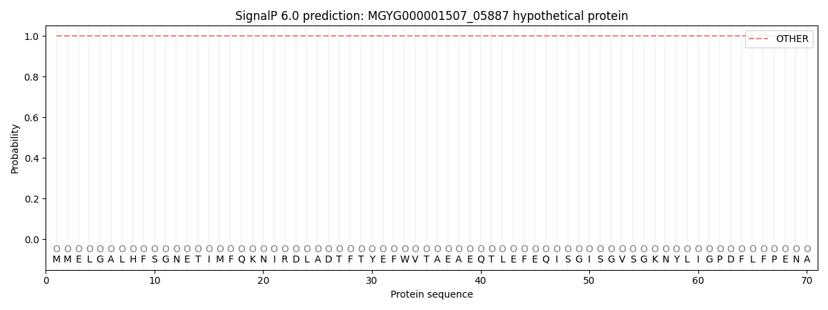
SignalP and Lipop Annotations help
This protein is predicted as OTHER
| Other | SP_Sec_SPI | LIPO_Sec_SPII | TAT_Tat_SPI | TATLIP_Sec_SPII | PILIN_Sec_SPIII |
|---|---|---|---|---|---|
| 1.000042 | 0.000012 | 0.000000 | 0.000000 | 0.000000 | 0.000000 |