You are browsing environment: HUMAN GUT
CAZyme Information: MGYG000001512_02357
You are here: Home > Sequence: MGYG000001512_02357
Basic Information |
Genomic context |
Full Sequence |
Enzyme annotations |
CAZy signature domains |
CDD domains |
CAZyme hits |
PDB hits |
Swiss-Prot hits |
SignalP and Lipop annotations |
TMHMM annotations
Basic Information help
| Species | Coprobacter secundus | |||||||||||
|---|---|---|---|---|---|---|---|---|---|---|---|---|
| Lineage | Bacteria; Bacteroidota; Bacteroidia; Bacteroidales; Coprobacteraceae; Coprobacter; Coprobacter secundus | |||||||||||
| CAZyme ID | MGYG000001512_02357 | |||||||||||
| CAZy Family | GT4 | |||||||||||
| CAZyme Description | D-inositol-3-phosphate glycosyltransferase | |||||||||||
| CAZyme Property |
|
|||||||||||
| Genome Property |
|
|||||||||||
| Gene Location | Start: 414620; End: 415885 Strand: - | |||||||||||
CDD Domains download full data without filtering help
| Cdd ID | Domain | E-Value | qStart | qEnd | sStart | sEnd | Domain Description |
|---|---|---|---|---|---|---|---|
| cd03809 | GT4_MtfB-like | 3.23e-106 | 21 | 384 | 1 | 358 | glycosyltransferases MtfB, WbpX, and similar proteins. This family is most closely related to the GT4 family of glycosyltransferases. MtfB (mannosyltransferase B) in E. coli has been shown to direct the growth of the O9-specific polysaccharide chain. It transfers two mannoses into the position 3 of the previously synthesized polysaccharide. |
| cd03801 | GT4_PimA-like | 9.98e-52 | 35 | 391 | 15 | 366 | phosphatidyl-myo-inositol mannosyltransferase. This family is most closely related to the GT4 family of glycosyltransferases and named after PimA in Propionibacterium freudenreichii, which is involved in the biosynthesis of phosphatidyl-myo-inositol mannosides (PIM) which are early precursors in the biosynthesis of lipomannans (LM) and lipoarabinomannans (LAM), and catalyzes the addition of a mannosyl residue from GDP-D-mannose (GDP-Man) to the position 2 of the carrier lipid phosphatidyl-myo-inositol (PI) to generate a phosphatidyl-myo-inositol bearing an alpha-1,2-linked mannose residue (PIM1). Glycosyltransferases catalyze the transfer of sugar moieties from activated donor molecules to specific acceptor molecules, forming glycosidic bonds. The acceptor molecule can be a lipid, a protein, a heterocyclic compound, or another carbohydrate residue. This group of glycosyltransferases is most closely related to the previously defined glycosyltransferase family 1 (GT1). The members of this family may transfer UDP, ADP, GDP, or CMP linked sugars. The diverse enzymatic activities among members of this family reflect a wide range of biological functions. The protein structure available for this family has the GTB topology, one of the two protein topologies observed for nucleotide-sugar-dependent glycosyltransferases. GTB proteins have distinct N- and C- terminal domains each containing a typical Rossmann fold. The two domains have high structural homology despite minimal sequence homology. The large cleft that separates the two domains includes the catalytic center and permits a high degree of flexibility. The members of this family are found mainly in certain bacteria and archaea. |
| COG0438 | RfaB | 5.73e-41 | 20 | 392 | 1 | 376 | Glycosyltransferase involved in cell wall bisynthesis [Cell wall/membrane/envelope biogenesis]. |
| cd03811 | GT4_GT28_WabH-like | 8.33e-31 | 41 | 379 | 16 | 351 | family 4 and family 28 glycosyltransferases similar to Klebsiella WabH. This family is most closely related to the GT1 family of glycosyltransferases. WabH in Klebsiella pneumoniae has been shown to transfer a GlcNAc residue from UDP-GlcNAc onto the acceptor GalUA residue in the cellular outer core. |
| cd03794 | GT4_WbuB-like | 4.25e-30 | 124 | 384 | 120 | 388 | Escherichia coli WbuB and similar proteins. This family is most closely related to the GT1 family of glycosyltransferases. WbuB in E. coli is involved in the biosynthesis of the O26 O-antigen. It has been proposed to function as an N-acetyl-L-fucosamine (L-FucNAc) transferase. |
CAZyme Hits help
| Hit ID | E-Value | Query Start | Query End | Hit Start | Hit End |
|---|---|---|---|---|---|
| BCI64068.1 | 7.60e-307 | 1 | 421 | 1 | 421 |
| AHF12636.1 | 7.83e-171 | 20 | 408 | 1 | 388 |
| QNL40319.1 | 9.42e-150 | 20 | 392 | 1 | 374 |
| QGT72872.1 | 3.81e-149 | 20 | 392 | 1 | 374 |
| SCV07516.1 | 1.09e-148 | 20 | 392 | 1 | 374 |
PDB Hits download full data without filtering help
| Hit ID | E-Value | Query Start | Query End | Hit Start | Hit End | Description |
|---|---|---|---|---|---|---|
| 3C4Q_A | 1.17e-13 | 165 | 398 | 165 | 410 | Structureof the retaining glycosyltransferase MshA : The first step in mycothiol biosynthesis. Organism : Corynebacterium glutamicum- Complex with UDP [Corynebacterium glutamicum],3C4Q_B Structure of the retaining glycosyltransferase MshA : The first step in mycothiol biosynthesis. Organism : Corynebacterium glutamicum- Complex with UDP [Corynebacterium glutamicum],3C4V_A Structure of the retaining glycosyltransferase MshA:The first step in mycothiol biosynthesis. Organism: Corynebacterium glutamicum : Complex with UDP and 1L-INS-1-P. [Corynebacterium glutamicum],3C4V_B Structure of the retaining glycosyltransferase MshA:The first step in mycothiol biosynthesis. Organism: Corynebacterium glutamicum : Complex with UDP and 1L-INS-1-P. [Corynebacterium glutamicum] |
| 3C48_A | 1.22e-13 | 165 | 398 | 185 | 430 | Structureof the retaining glycosyltransferase MshA: The first step in mycothiol biosynthesis. Organism: Corynebacterium glutamicum- APO (OPEN) structure. [Corynebacterium glutamicum],3C48_B Structure of the retaining glycosyltransferase MshA: The first step in mycothiol biosynthesis. Organism: Corynebacterium glutamicum- APO (OPEN) structure. [Corynebacterium glutamicum] |
| 5I45_A | 3.60e-07 | 219 | 395 | 26 | 214 | 1.35Angstrom Crystal Structure of C-terminal Domain of Glycosyl Transferase Group 1 Family Protein (LpcC) from Francisella tularensis. [Francisella tularensis subsp. tularensis SCHU S4] |
| 2R60_A | 1.49e-06 | 204 | 400 | 265 | 462 | Structureof apo Sucrose Phosphate Synthase (SPS) of Halothermothrix orenii [Halothermothrix orenii],2R66_A Complex Structure of Sucrose Phosphate Synthase (SPS)-F6P of Halothermothrix orenii [Halothermothrix orenii H 168],2R68_A Complex Structure of Sucrose Phosphate Synthase (SPS)-S6P of Halothermothrix orenii [Halothermothrix orenii H 168] |
Swiss-Prot Hits download full data without filtering help
| Hit ID | E-Value | Query Start | Query End | Hit Start | Hit End | Description |
|---|---|---|---|---|---|---|
| P26402 | 5.14e-24 | 20 | 377 | 1 | 336 | Protein RfbU OS=Salmonella typhimurium (strain LT2 / SGSC1412 / ATCC 700720) OX=99287 GN=rfbU PE=3 SV=1 |
| Q65CC7 | 2.76e-15 | 127 | 366 | 110 | 356 | Alpha-D-kanosaminyltransferase OS=Streptomyces kanamyceticus OX=1967 GN=kanE PE=1 SV=1 |
| A4QB40 | 6.22e-13 | 165 | 398 | 165 | 410 | D-inositol 3-phosphate glycosyltransferase OS=Corynebacterium glutamicum (strain R) OX=340322 GN=mshA PE=3 SV=1 |
| Q8NTA6 | 6.22e-13 | 165 | 398 | 165 | 410 | D-inositol 3-phosphate glycosyltransferase OS=Corynebacterium glutamicum (strain ATCC 13032 / DSM 20300 / BCRC 11384 / JCM 1318 / LMG 3730 / NCIMB 10025) OX=196627 GN=mshA PE=1 SV=1 |
| A0QQZ8 | 6.60e-12 | 165 | 357 | 176 | 372 | D-inositol 3-phosphate glycosyltransferase OS=Mycolicibacterium smegmatis (strain ATCC 700084 / mc(2)155) OX=246196 GN=mshA PE=1 SV=1 |
SignalP and Lipop Annotations help
This protein is predicted as OTHER
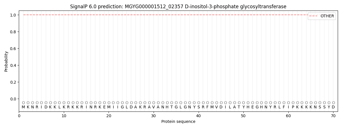
| Other | SP_Sec_SPI | LIPO_Sec_SPII | TAT_Tat_SPI | TATLIP_Sec_SPII | PILIN_Sec_SPIII |
|---|---|---|---|---|---|
| 1.000048 | 0.000000 | 0.000000 | 0.000000 | 0.000000 | 0.000000 |