You are browsing environment: HUMAN GUT
CAZyme Information: MGYG000001678_00524
You are here: Home > Sequence: MGYG000001678_00524
Basic Information |
Genomic context |
Full Sequence |
Enzyme annotations |
CAZy signature domains |
CDD domains |
CAZyme hits |
PDB hits |
Swiss-Prot hits |
SignalP and Lipop annotations |
TMHMM annotations
Basic Information help
| Species | ||||||||||||
|---|---|---|---|---|---|---|---|---|---|---|---|---|
| Lineage | Bacteria; Cyanobacteria; Vampirovibrionia; Gastranaerophilales; Gastranaerophilaceae; Zag1; | |||||||||||
| CAZyme ID | MGYG000001678_00524 | |||||||||||
| CAZy Family | GT2 | |||||||||||
| CAZyme Description | 2,3,4,5-tetrahydropyridine-2,6-dicarboxylate N-acetyltransferase | |||||||||||
| CAZyme Property |
|
|||||||||||
| Genome Property |
|
|||||||||||
| Gene Location | Start: 53127; End: 54665 Strand: + | |||||||||||
CAZyme Signature Domains help
| Family | Start | End | Evalue | family coverage |
|---|---|---|---|---|
| GT2 | 7 | 118 | 4.1e-17 | 0.6294117647058823 |
CDD Domains download full data without filtering help
| Cdd ID | Domain | E-Value | qStart | qEnd | sStart | sEnd | Domain Description |
|---|---|---|---|---|---|---|---|
| cd03349 | LbH_XAT | 1.12e-52 | 338 | 471 | 1 | 137 | Xenobiotic acyltransferase (XAT): The XAT class of hexapeptide acyltransferases is composed of a large number of microbial enzymes that catalyze the CoA-dependent acetylation of a variety of hydroxyl-bearing acceptors such as chloramphenicol and streptogramin, among others. Members of this class of enzymes include Enterococcus faecium streptogramin A acetyltransferase and Pseudomonas aeruginosa chloramphenicol acetyltransferase. They contain repeated copies of a six-residue hexapeptide repeat sequence motif (X-[STAV]-X-[LIV]-[GAED]-X) and adopt a left-handed parallel beta helix (LbH) structure. The active enzyme is a trimer with CoA and substrate binding sites at the interface of two separate LbH subunits. XATs are implicated in inactivating xenobiotics leading to xenobiotic resistance in patients. |
| cd04647 | LbH_MAT_like | 6.80e-32 | 338 | 458 | 1 | 109 | Maltose O-acyltransferase (MAT)-like: This family is composed of maltose O-acetyltransferase, galactoside O-acetyltransferase (GAT), xenobiotic acyltransferase (XAT) and similar proteins. MAT and GAT catalyze the CoA-dependent acetylation of the 6-hydroxyl group of their respective sugar substrates. MAT acetylates maltose and glucose exclusively while GAT specifically acetylates galactopyranosides. XAT catalyzes the CoA-dependent acetylation of a variety of hydroxyl-bearing acceptors such as chloramphenicol and streptogramin, among others. XATs are implicated in inactivating xenobiotics leading to xenobiotic resistance in patients. Members of this family contain a a left-handed parallel beta-helix (LbH) domain with at least 5 turns, each containing three imperfect tandem repeats of a hexapeptide repeat motif (X-[STAV]-X-[LIV]-[GAED]-X). They are trimeric in their active form. |
| cd03357 | LbH_MAT_GAT | 8.38e-26 | 337 | 456 | 61 | 167 | Maltose O-acetyltransferase (MAT) and Galactoside O-acetyltransferase (GAT): MAT and GAT catalyze the CoA-dependent acetylation of the 6-hydroxyl group of their respective sugar substrates. MAT acetylates maltose and glucose exclusively at the C6 position of the nonreducing end glucosyl moiety. GAT specifically acetylates galactopyranosides. Furthermore, MAT shows higher affinity toward artificial substrates containing an alkyl or hydrophobic chain as well as a glucosyl unit. Active MAT and GAT are homotrimers, with each subunit consisting of an N-terminal alpha-helical region and a C-terminal left-handed parallel alpha-helix (LbH) subdomain with 6 turns, each containing three imperfect tandem repeats of a hexapeptide repeat motif (X-[STAV]-X-[LIV]-[GAED]-X). |
| COG0110 | WbbJ | 1.44e-24 | 328 | 471 | 57 | 188 | Acetyltransferase (isoleucine patch superfamily) [General function prediction only]. |
| cd05825 | LbH_wcaF_like | 1.53e-21 | 356 | 458 | 19 | 107 | wcaF-like: This group is composed of the protein product of the E. coli wcaF gene and similar proteins. WcaF is part of the gene cluster responsible for the biosynthesis of the extracellular polysaccharide colanic acid. The wcaF protein is predicted to contain a left-handed parallel beta-helix (LbH) domain encoded by imperfect tandem repeats of a hexapeptide repeat motif (X-[STAV]-X-[LIV]-[GAED]-X). Proteins containing hexapeptide repeats are often enzymes showing acyltransferase activity. Many are trimeric in their active forms. |
CAZyme Hits help
| Hit ID | E-Value | Query Start | Query End | Hit Start | Hit End |
|---|---|---|---|---|---|
| AEF84575.1 | 6.44e-50 | 4 | 503 | 31 | 573 |
| SDT46839.1 | 4.32e-40 | 6 | 298 | 4 | 299 |
| AMR32929.1 | 1.29e-37 | 1 | 250 | 4 | 255 |
| QKJ31486.1 | 2.08e-35 | 6 | 264 | 4 | 258 |
| AUS95090.1 | 5.37e-25 | 2 | 254 | 419 | 666 |
PDB Hits download full data without filtering help
| Hit ID | E-Value | Query Start | Query End | Hit Start | Hit End | Description |
|---|---|---|---|---|---|---|
| 6MFK_A | 6.78e-24 | 361 | 492 | 65 | 211 | ChainA, Chloramphenicol acetyltransferase [Elizabethkingia anophelis] |
| 1KHR_A | 1.02e-20 | 338 | 493 | 29 | 199 | CrystalStructure of Vat(D) in Complex with Virginiamycin and Coenzyme A [Enterococcus faecium],1KHR_B Crystal Structure of Vat(D) in Complex with Virginiamycin and Coenzyme A [Enterococcus faecium],1KHR_C Crystal Structure of Vat(D) in Complex with Virginiamycin and Coenzyme A [Enterococcus faecium],1KHR_D Crystal Structure of Vat(D) in Complex with Virginiamycin and Coenzyme A [Enterococcus faecium],1KHR_E Crystal Structure of Vat(D) in Complex with Virginiamycin and Coenzyme A [Enterococcus faecium],1KHR_F Crystal Structure of Vat(D) in Complex with Virginiamycin and Coenzyme A [Enterococcus faecium],1KK4_A Crystal Structure of Vat(D) in Complex with Acetyl-CoA [Enterococcus faecium],1KK4_B Crystal Structure of Vat(D) in Complex with Acetyl-CoA [Enterococcus faecium],1KK4_C Crystal Structure of Vat(D) in Complex with Acetyl-CoA [Enterococcus faecium],1KK4_D Crystal Structure of Vat(D) in Complex with Acetyl-CoA [Enterococcus faecium],1KK4_E Crystal Structure of Vat(D) in Complex with Acetyl-CoA [Enterococcus faecium],1KK4_F Crystal Structure of Vat(D) in Complex with Acetyl-CoA [Enterococcus faecium],1KK5_A Crystal Structure of Vat(D) (Form II) [Enterococcus faecium],1KK5_B Crystal Structure of Vat(D) (Form II) [Enterococcus faecium],1KK5_C Crystal Structure of Vat(D) (Form II) [Enterococcus faecium],1KK5_D Crystal Structure of Vat(D) (Form II) [Enterococcus faecium],1KK5_E Crystal Structure of Vat(D) (Form II) [Enterococcus faecium],1KK5_F Crystal Structure of Vat(D) (Form II) [Enterococcus faecium],1KK6_A Crystal Structure of Vat(D) (Form I) [Enterococcus faecium],1KK6_B Crystal Structure of Vat(D) (Form I) [Enterococcus faecium],1KK6_C Crystal Structure of Vat(D) (Form I) [Enterococcus faecium],1MR7_A Crystal Structure of Streptogramin A Acetyltransferase [Enterococcus faecium],1MR7_B Crystal Structure of Streptogramin A Acetyltransferase [Enterococcus faecium],1MR7_C Crystal Structure of Streptogramin A Acetyltransferase [Enterococcus faecium],1MR7_X Crystal Structure of Streptogramin A Acetyltransferase [Enterococcus faecium],1MR7_Y Crystal Structure of Streptogramin A Acetyltransferase [Enterococcus faecium],1MR7_Z Crystal Structure of Streptogramin A Acetyltransferase [Enterococcus faecium],1MRL_A Crystal structure of streptogramin A acetyltransferase with dalfopristin [Enterococcus faecium],1MRL_B Crystal structure of streptogramin A acetyltransferase with dalfopristin [Enterococcus faecium],1MRL_C Crystal structure of streptogramin A acetyltransferase with dalfopristin [Enterococcus faecium],3DHO_A Structure of Streptogramin Acetyltransferase in Complex with an Inhibitor [Enterococcus faecium],3DHO_B Structure of Streptogramin Acetyltransferase in Complex with an Inhibitor [Enterococcus faecium],3DHO_C Structure of Streptogramin Acetyltransferase in Complex with an Inhibitor [Enterococcus faecium],3DHO_D Structure of Streptogramin Acetyltransferase in Complex with an Inhibitor [Enterococcus faecium],3DHO_E Structure of Streptogramin Acetyltransferase in Complex with an Inhibitor [Enterococcus faecium],3DHO_F Structure of Streptogramin Acetyltransferase in Complex with an Inhibitor [Enterococcus faecium] |
| 3EEV_A | 6.05e-19 | 356 | 472 | 54 | 176 | CrystalStructure of Chloramphenicol Acetyltransferase VCA0300 from Vibrio cholerae O1 biovar eltor [Vibrio cholerae O1 biovar El Tor str. N16961],3EEV_B Crystal Structure of Chloramphenicol Acetyltransferase VCA0300 from Vibrio cholerae O1 biovar eltor [Vibrio cholerae O1 biovar El Tor str. N16961],3EEV_C Crystal Structure of Chloramphenicol Acetyltransferase VCA0300 from Vibrio cholerae O1 biovar eltor [Vibrio cholerae O1 biovar El Tor str. N16961],6PUA_A The 2.0 A Crystal Structure of the Type B Chloramphenicol Acetyltransferase from Vibrio cholerae [Vibrio cholerae O1 biovar El Tor str. N16961],6PUA_B The 2.0 A Crystal Structure of the Type B Chloramphenicol Acetyltransferase from Vibrio cholerae [Vibrio cholerae O1 biovar El Tor str. N16961],6PUA_C The 2.0 A Crystal Structure of the Type B Chloramphenicol Acetyltransferase from Vibrio cholerae [Vibrio cholerae O1 biovar El Tor str. N16961],6PUB_A Crystal Structure of the Type B Chloramphenicol Acetyltransferase from Vibrio cholerae in the Complex with Crystal Violet [Vibrio cholerae O1 biovar El Tor str. N16961],6U9C_A The 2.2 A Crystal Structure of the Type B Chloramphenicol Acetyltransferase from Vibrio cholerae in the complex with Acetyl CoA [Vibrio cholerae O1 biovar El Tor str. N16961],6U9C_B The 2.2 A Crystal Structure of the Type B Chloramphenicol Acetyltransferase from Vibrio cholerae in the complex with Acetyl CoA [Vibrio cholerae O1 biovar El Tor str. N16961],6U9C_C The 2.2 A Crystal Structure of the Type B Chloramphenicol Acetyltransferase from Vibrio cholerae in the complex with Acetyl CoA [Vibrio cholerae O1 biovar El Tor str. N16961] |
| 6PU9_A | 9.24e-18 | 361 | 470 | 59 | 174 | CrystalStructure of the Type B Chloramphenicol O-Acetyltransferase from Vibrio vulnificus [Vibrio vulnificus CMCP6],6PU9_B Crystal Structure of the Type B Chloramphenicol O-Acetyltransferase from Vibrio vulnificus [Vibrio vulnificus CMCP6],6PU9_C Crystal Structure of the Type B Chloramphenicol O-Acetyltransferase from Vibrio vulnificus [Vibrio vulnificus CMCP6] |
| 6PXA_A | 7.25e-17 | 361 | 471 | 71 | 184 | Thecrystal structure of chloramphenicol acetyltransferase-like protein from Vibrio fischeri ES114 in complex with taurocholic acid [Aliivibrio fischeri ES114],6PXA_B The crystal structure of chloramphenicol acetyltransferase-like protein from Vibrio fischeri ES114 in complex with taurocholic acid [Aliivibrio fischeri ES114],6PXA_C The crystal structure of chloramphenicol acetyltransferase-like protein from Vibrio fischeri ES114 in complex with taurocholic acid [Aliivibrio fischeri ES114],6PXA_D The crystal structure of chloramphenicol acetyltransferase-like protein from Vibrio fischeri ES114 in complex with taurocholic acid [Aliivibrio fischeri ES114],6PXA_E The crystal structure of chloramphenicol acetyltransferase-like protein from Vibrio fischeri ES114 in complex with taurocholic acid [Aliivibrio fischeri ES114],6PXA_F The crystal structure of chloramphenicol acetyltransferase-like protein from Vibrio fischeri ES114 in complex with taurocholic acid [Aliivibrio fischeri ES114],6PXA_G The crystal structure of chloramphenicol acetyltransferase-like protein from Vibrio fischeri ES114 in complex with taurocholic acid [Aliivibrio fischeri ES114],6PXA_H The crystal structure of chloramphenicol acetyltransferase-like protein from Vibrio fischeri ES114 in complex with taurocholic acid [Aliivibrio fischeri ES114],6PXA_I The crystal structure of chloramphenicol acetyltransferase-like protein from Vibrio fischeri ES114 in complex with taurocholic acid [Aliivibrio fischeri ES114],6PXA_J The crystal structure of chloramphenicol acetyltransferase-like protein from Vibrio fischeri ES114 in complex with taurocholic acid [Aliivibrio fischeri ES114],6PXA_K The crystal structure of chloramphenicol acetyltransferase-like protein from Vibrio fischeri ES114 in complex with taurocholic acid [Aliivibrio fischeri ES114],6PXA_L The crystal structure of chloramphenicol acetyltransferase-like protein from Vibrio fischeri ES114 in complex with taurocholic acid [Aliivibrio fischeri ES114] |
Swiss-Prot Hits download full data without filtering help
| Hit ID | E-Value | Query Start | Query End | Hit Start | Hit End | Description |
|---|---|---|---|---|---|---|
| P26840 | 2.33e-21 | 361 | 490 | 24 | 160 | Probable macrolide acetyltransferase (Fragment) OS=Lysinibacillus sphaericus OX=1421 PE=3 SV=1 |
| P23364 | 5.59e-20 | 336 | 492 | 20 | 202 | Chloramphenicol acetyltransferase OS=Agrobacterium fabrum (strain C58 / ATCC 33970) OX=176299 GN=cat PE=3 SV=1 |
| P50870 | 5.59e-20 | 338 | 493 | 29 | 199 | Streptogramin A acetyltransferase OS=Enterococcus faecium OX=1352 GN=vatD PE=1 SV=1 |
| P26838 | 1.06e-19 | 356 | 471 | 52 | 174 | Chloramphenicol acetyltransferase OS=Escherichia coli OX=562 GN=catB2 PE=3 SV=2 |
| P50868 | 2.69e-19 | 356 | 472 | 52 | 175 | Chloramphenicol acetyltransferase OS=Klebsiella aerogenes OX=548 GN=catB4 PE=3 SV=1 |
SignalP and Lipop Annotations help
This protein is predicted as OTHER
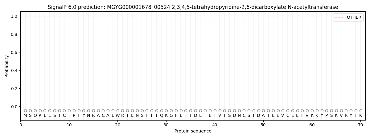
| Other | SP_Sec_SPI | LIPO_Sec_SPII | TAT_Tat_SPI | TATLIP_Sec_SPII | PILIN_Sec_SPIII |
|---|---|---|---|---|---|
| 1.000045 | 0.000000 | 0.000000 | 0.000000 | 0.000000 | 0.000000 |