You are browsing environment: HUMAN GUT
CAZyme Information: MGYG000001688_02351
You are here: Home > Sequence: MGYG000001688_02351
Basic Information |
Genomic context |
Full Sequence |
Enzyme annotations |
CAZy signature domains |
CDD domains |
CAZyme hits |
PDB hits |
Swiss-Prot hits |
SignalP and Lipop annotations |
TMHMM annotations
Basic Information help
| Species | Hungatella_A hathewayi_A | |||||||||||
|---|---|---|---|---|---|---|---|---|---|---|---|---|
| Lineage | Bacteria; Firmicutes_A; Clostridia; Lachnospirales; Lachnospiraceae; Hungatella_A; Hungatella_A hathewayi_A | |||||||||||
| CAZyme ID | MGYG000001688_02351 | |||||||||||
| CAZy Family | GT2 | |||||||||||
| CAZyme Description | hypothetical protein | |||||||||||
| CAZyme Property |
|
|||||||||||
| Genome Property |
|
|||||||||||
| Gene Location | Start: 30370; End: 31236 Strand: + | |||||||||||
CAZyme Signature Domains help
| Family | Start | End | Evalue | family coverage |
|---|---|---|---|---|
| GT2 | 8 | 173 | 1.5e-29 | 0.9764705882352941 |
CDD Domains download full data without filtering help
| Cdd ID | Domain | E-Value | qStart | qEnd | sStart | sEnd | Domain Description |
|---|---|---|---|---|---|---|---|
| COG1216 | GT2 | 2.15e-63 | 5 | 286 | 4 | 291 | Glycosyltransferase, GT2 family [Carbohydrate transport and metabolism]. |
| cd04186 | GT_2_like_c | 9.29e-59 | 8 | 213 | 1 | 166 | Subfamily of Glycosyltransferase Family GT2 of unknown function. GT-2 includes diverse families of glycosyltransferases with a common GT-A type structural fold, which has two tightly associated beta/alpha/beta domains that tend to form a continuous central sheet of at least eight beta-strands. These are enzymes that catalyze the transfer of sugar moieties from activated donor molecules to specific acceptor molecules, forming glycosidic bonds. Glycosyltransferases have been classified into more than 90 distinct sequence based families. |
| cd04185 | GT_2_like_b | 5.90e-36 | 9 | 236 | 2 | 202 | Subfamily of Glycosyltransferase Family GT2 of unknown function. GT-2 includes diverse families of glycosyltransferases with a common GT-A type structural fold, which has two tightly associated beta/alpha/beta domains that tend to form a continuous central sheet of at least eight beta-strands. These are enzymes that catalyze the transfer of sugar moieties from activated donor molecules to specific acceptor molecules, forming glycosidic bonds. Glycosyltransferases have been classified into more than 90 distinct sequence based families. |
| pfam00535 | Glycos_transf_2 | 2.07e-27 | 8 | 171 | 2 | 164 | Glycosyl transferase family 2. Diverse family, transferring sugar from UDP-glucose, UDP-N-acetyl- galactosamine, GDP-mannose or CDP-abequose, to a range of substrates including cellulose, dolichol phosphate and teichoic acids. |
| cd02526 | GT2_RfbF_like | 1.67e-26 | 9 | 237 | 2 | 237 | RfbF is a putative dTDP-rhamnosyl transferase. Shigella flexneri RfbF protein is a putative dTDP-rhamnosyl transferase. dTDP rhamnosyl transferases of Shigella flexneri add rhamnose sugars to N-acetyl-glucosamine in the O-antigen tetrasaccharide repeat. Lipopolysaccharide O antigens are important virulence determinants for many bacteria. The variations of sugar composition, the sequence of the sugars and the linkages in the O antigen provide structural diversity of the O antigen. |
CAZyme Hits help
| Hit ID | E-Value | Query Start | Query End | Hit Start | Hit End |
|---|---|---|---|---|---|
| ABY95272.1 | 1.34e-65 | 5 | 288 | 8 | 303 |
| ADV80219.1 | 1.34e-65 | 5 | 288 | 8 | 303 |
| AEM74867.1 | 3.28e-65 | 1 | 284 | 1 | 296 |
| ADD01946.1 | 3.19e-64 | 1 | 288 | 1 | 300 |
| QSW21431.1 | 6.58e-61 | 5 | 286 | 3 | 294 |
PDB Hits download full data without filtering help
| Hit ID | E-Value | Query Start | Query End | Hit Start | Hit End | Description |
|---|---|---|---|---|---|---|
| 5HEA_A | 2.82e-09 | 4 | 108 | 5 | 105 | CgTstructure in hexamer [Streptococcus parasanguinis FW213],5HEA_B CgT structure in hexamer [Streptococcus parasanguinis FW213],5HEA_C CgT structure in hexamer [Streptococcus parasanguinis FW213],5HEC_A CgT structure in dimer [Streptococcus parasanguinis FW213],5HEC_B CgT structure in dimer [Streptococcus parasanguinis FW213] |
| 3L7I_A | 9.64e-09 | 5 | 172 | 3 | 165 | Structureof the Wall Teichoic Acid Polymerase TagF [Staphylococcus epidermidis RP62A],3L7I_B Structure of the Wall Teichoic Acid Polymerase TagF [Staphylococcus epidermidis RP62A],3L7I_C Structure of the Wall Teichoic Acid Polymerase TagF [Staphylococcus epidermidis RP62A],3L7I_D Structure of the Wall Teichoic Acid Polymerase TagF [Staphylococcus epidermidis RP62A] |
| 3L7J_A | 9.64e-09 | 5 | 172 | 3 | 165 | ChainA, Teichoic acid biosynthesis protein F [Staphylococcus epidermidis RP62A],3L7J_B Chain B, Teichoic acid biosynthesis protein F [Staphylococcus epidermidis RP62A],3L7J_C Chain C, Teichoic acid biosynthesis protein F [Staphylococcus epidermidis RP62A],3L7J_D Chain D, Teichoic acid biosynthesis protein F [Staphylococcus epidermidis RP62A],3L7K_A Chain A, Teichoic acid biosynthesis protein F [Staphylococcus epidermidis RP62A],3L7K_B Chain B, Teichoic acid biosynthesis protein F [Staphylococcus epidermidis RP62A],3L7K_C Chain C, Teichoic acid biosynthesis protein F [Staphylococcus epidermidis RP62A],3L7K_D Chain D, Teichoic acid biosynthesis protein F [Staphylococcus epidermidis RP62A],3L7L_A Chain A, Teichoic acid biosynthesis protein F [Staphylococcus epidermidis RP62A],3L7L_B Chain B, Teichoic acid biosynthesis protein F [Staphylococcus epidermidis RP62A],3L7L_C Chain C, Teichoic acid biosynthesis protein F [Staphylococcus epidermidis RP62A],3L7L_D Chain D, Teichoic acid biosynthesis protein F [Staphylococcus epidermidis RP62A] |
| 3L7M_A | 9.64e-09 | 5 | 172 | 3 | 165 | ChainA, Teichoic acid biosynthesis protein F [Staphylococcus epidermidis RP62A],3L7M_B Chain B, Teichoic acid biosynthesis protein F [Staphylococcus epidermidis RP62A],3L7M_C Chain C, Teichoic acid biosynthesis protein F [Staphylococcus epidermidis RP62A],3L7M_D Chain D, Teichoic acid biosynthesis protein F [Staphylococcus epidermidis RP62A] |
| 2Z87_A | 2.95e-06 | 6 | 114 | 376 | 480 | Crystalstructure of chondroitin polymerase from Escherichia coli strain K4 (K4CP) complexed with UDP-GalNAc and UDP [Escherichia coli],2Z87_B Crystal structure of chondroitin polymerase from Escherichia coli strain K4 (K4CP) complexed with UDP-GalNAc and UDP [Escherichia coli] |
Swiss-Prot Hits download full data without filtering help
| Hit ID | E-Value | Query Start | Query End | Hit Start | Hit End | Description |
|---|---|---|---|---|---|---|
| D4GU63 | 1.37e-17 | 8 | 234 | 22 | 246 | Low-salt glycan biosynthesis hexosyltransferase Agl10 OS=Haloferax volcanii (strain ATCC 29605 / DSM 3757 / JCM 8879 / NBRC 14742 / NCIMB 2012 / VKM B-1768 / DS2) OX=309800 GN=agl10 PE=3 SV=1 |
| P37782 | 1.09e-13 | 6 | 239 | 5 | 241 | dTDP-rhamnosyl transferase RfbF OS=Shigella flexneri OX=623 GN=rfbF PE=3 SV=2 |
| A0A0H2UR96 | 2.65e-09 | 4 | 126 | 3 | 122 | Glycosyltransferase GlyG OS=Streptococcus pneumoniae serotype 4 (strain ATCC BAA-334 / TIGR4) OX=170187 GN=glyG PE=1 SV=1 |
| Q5HLM5 | 5.26e-08 | 5 | 172 | 3 | 165 | Teichoic acid poly(glycerol phosphate) polymerase OS=Staphylococcus epidermidis (strain ATCC 35984 / RP62A) OX=176279 GN=tagF PE=1 SV=1 |
| D4GYH2 | 7.24e-07 | 6 | 216 | 9 | 212 | Glycosyltransferase AglI OS=Haloferax volcanii (strain ATCC 29605 / DSM 3757 / JCM 8879 / NBRC 14742 / NCIMB 2012 / VKM B-1768 / DS2) OX=309800 GN=aglI PE=1 SV=1 |
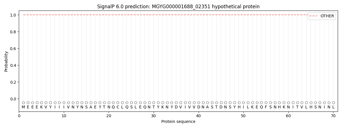
SignalP and Lipop Annotations help
This protein is predicted as OTHER
| Other | SP_Sec_SPI | LIPO_Sec_SPII | TAT_Tat_SPI | TATLIP_Sec_SPII | PILIN_Sec_SPIII |
|---|---|---|---|---|---|
| 1.000059 | 0.000000 | 0.000000 | 0.000000 | 0.000000 | 0.000000 |