You are browsing environment: HUMAN GUT
CAZyme Information: MGYG000001705_00099
You are here: Home > Sequence: MGYG000001705_00099
Basic Information |
Genomic context |
Full Sequence |
Enzyme annotations |
CAZy signature domains |
CDD domains |
CAZyme hits |
PDB hits |
Swiss-Prot hits |
SignalP and Lipop annotations |
TMHMM annotations
Basic Information help
| Species | Citrobacter portucalensis | |||||||||||
|---|---|---|---|---|---|---|---|---|---|---|---|---|
| Lineage | Bacteria; Proteobacteria; Gammaproteobacteria; Enterobacterales; Enterobacteriaceae; Citrobacter; Citrobacter portucalensis | |||||||||||
| CAZyme ID | MGYG000001705_00099 | |||||||||||
| CAZy Family | GT4 | |||||||||||
| CAZyme Description | Lipopolysaccharide core biosynthesis protein RfaG | |||||||||||
| CAZyme Property |
|
|||||||||||
| Genome Property |
|
|||||||||||
| Gene Location | Start: 98730; End: 99854 Strand: + | |||||||||||
CDD Domains download full data without filtering help
| Cdd ID | Domain | E-Value | qStart | qEnd | sStart | sEnd | Domain Description |
|---|---|---|---|---|---|---|---|
| cd03801 | GT4_PimA-like | 3.40e-44 | 2 | 369 | 1 | 362 | phosphatidyl-myo-inositol mannosyltransferase. This family is most closely related to the GT4 family of glycosyltransferases and named after PimA in Propionibacterium freudenreichii, which is involved in the biosynthesis of phosphatidyl-myo-inositol mannosides (PIM) which are early precursors in the biosynthesis of lipomannans (LM) and lipoarabinomannans (LAM), and catalyzes the addition of a mannosyl residue from GDP-D-mannose (GDP-Man) to the position 2 of the carrier lipid phosphatidyl-myo-inositol (PI) to generate a phosphatidyl-myo-inositol bearing an alpha-1,2-linked mannose residue (PIM1). Glycosyltransferases catalyze the transfer of sugar moieties from activated donor molecules to specific acceptor molecules, forming glycosidic bonds. The acceptor molecule can be a lipid, a protein, a heterocyclic compound, or another carbohydrate residue. This group of glycosyltransferases is most closely related to the previously defined glycosyltransferase family 1 (GT1). The members of this family may transfer UDP, ADP, GDP, or CMP linked sugars. The diverse enzymatic activities among members of this family reflect a wide range of biological functions. The protein structure available for this family has the GTB topology, one of the two protein topologies observed for nucleotide-sugar-dependent glycosyltransferases. GTB proteins have distinct N- and C- terminal domains each containing a typical Rossmann fold. The two domains have high structural homology despite minimal sequence homology. The large cleft that separates the two domains includes the catalytic center and permits a high degree of flexibility. The members of this family are found mainly in certain bacteria and archaea. |
| pfam00534 | Glycos_transf_1 | 1.38e-31 | 195 | 351 | 1 | 158 | Glycosyl transferases group 1. Mutations in this domain of PIGA lead to disease (Paroxysmal Nocturnal haemoglobinuria). Members of this family transfer activated sugars to a variety of substrates, including glycogen, Fructose-6-phosphate and lipopolysaccharides. Members of this family transfer UDP, ADP, GDP or CMP linked sugars. The eukaryotic glycogen synthases may be distant members of this family. |
| COG0438 | RfaB | 4.98e-28 | 1 | 369 | 1 | 371 | Glycosyltransferase involved in cell wall bisynthesis [Cell wall/membrane/envelope biogenesis]. |
| cd03800 | GT4_sucrose_synthase | 1.85e-23 | 144 | 350 | 171 | 381 | sucrose-phosphate synthase and similar proteins. This family is most closely related to the GT4 family of glycosyltransferases. The sucrose-phosphate synthases in this family may be unique to plants and photosynthetic bacteria. This enzyme catalyzes the synthesis of sucrose 6-phosphate from fructose 6-phosphate and uridine 5'-diphosphate-glucose, a key regulatory step of sucrose metabolism. The activity of this enzyme is regulated by phosphorylation and moderated by the concentration of various metabolites and light. |
| cd03811 | GT4_GT28_WabH-like | 4.01e-20 | 2 | 319 | 1 | 311 | family 4 and family 28 glycosyltransferases similar to Klebsiella WabH. This family is most closely related to the GT1 family of glycosyltransferases. WabH in Klebsiella pneumoniae has been shown to transfer a GlcNAc residue from UDP-GlcNAc onto the acceptor GalUA residue in the cellular outer core. |
CAZyme Hits help
| Hit ID | E-Value | Query Start | Query End | Hit Start | Hit End |
|---|---|---|---|---|---|
| QCC99804.1 | 5.40e-274 | 1 | 374 | 1 | 374 |
| AWV24832.1 | 6.30e-273 | 1 | 374 | 1 | 374 |
| AUV41557.1 | 1.81e-272 | 1 | 374 | 1 | 374 |
| QNM18623.1 | 4.25e-271 | 1 | 374 | 1 | 374 |
| QNM24086.1 | 4.25e-271 | 1 | 374 | 1 | 374 |
PDB Hits download full data without filtering help
| Hit ID | E-Value | Query Start | Query End | Hit Start | Hit End | Description |
|---|---|---|---|---|---|---|
| 2IW1_A | 8.62e-187 | 1 | 370 | 1 | 370 | CrystalStructure of WaaG, a glycosyltransferase involved in lipopolysaccharide biosynthesis [Escherichia coli str. K-12 substr. W3110] |
| 2IV7_A | 1.10e-182 | 3 | 370 | 3 | 370 | CrystalStructure of WaaG, a glycosyltransferase involved in lipopolysaccharide biosynthesis [Escherichia coli str. K-12 substr. W3110] |
| 2N58_A | 1.07e-08 | 103 | 132 | 1 | 30 | Structureof an N-terminal membrane-anchoring region of the glycosyltransferase WaaG [Escherichia coli K-12] |
| 2JJM_A | 9.78e-06 | 182 | 315 | 195 | 329 | CrystalStructure of a family GT4 glycosyltransferase from Bacillus anthracis ORF BA1558. [Bacillus anthracis str. Ames],2JJM_B Crystal Structure of a family GT4 glycosyltransferase from Bacillus anthracis ORF BA1558. [Bacillus anthracis str. Ames],2JJM_C Crystal Structure of a family GT4 glycosyltransferase from Bacillus anthracis ORF BA1558. [Bacillus anthracis str. Ames],2JJM_D Crystal Structure of a family GT4 glycosyltransferase from Bacillus anthracis ORF BA1558. [Bacillus anthracis str. Ames],2JJM_E Crystal Structure of a family GT4 glycosyltransferase from Bacillus anthracis ORF BA1558. [Bacillus anthracis str. Ames],2JJM_F Crystal Structure of a family GT4 glycosyltransferase from Bacillus anthracis ORF BA1558. [Bacillus anthracis str. Ames],2JJM_G Crystal Structure of a family GT4 glycosyltransferase from Bacillus anthracis ORF BA1558. [Bacillus anthracis str. Ames],2JJM_H Crystal Structure of a family GT4 glycosyltransferase from Bacillus anthracis ORF BA1558. [Bacillus anthracis str. Ames],2JJM_I Crystal Structure of a family GT4 glycosyltransferase from Bacillus anthracis ORF BA1558. [Bacillus anthracis str. Ames],2JJM_J Crystal Structure of a family GT4 glycosyltransferase from Bacillus anthracis ORF BA1558. [Bacillus anthracis str. Ames],2JJM_K Crystal Structure of a family GT4 glycosyltransferase from Bacillus anthracis ORF BA1558. [Bacillus anthracis str. Ames],2JJM_L Crystal Structure of a family GT4 glycosyltransferase from Bacillus anthracis ORF BA1558. [Bacillus anthracis str. Ames] |
Swiss-Prot Hits download full data without filtering help
| Hit ID | E-Value | Query Start | Query End | Hit Start | Hit End | Description |
|---|---|---|---|---|---|---|
| P25740 | 4.72e-186 | 1 | 370 | 1 | 370 | Lipopolysaccharide core biosynthesis protein RfaG OS=Escherichia coli (strain K12) OX=83333 GN=rfaG PE=1 SV=1 |
| D7C367 | 1.18e-08 | 135 | 319 | 200 | 392 | D-inositol 3-phosphate glycosyltransferase OS=Streptomyces bingchenggensis (strain BCW-1) OX=749414 GN=mshA PE=3 SV=1 |
| C9ZH13 | 6.42e-08 | 136 | 318 | 190 | 380 | D-inositol 3-phosphate glycosyltransferase OS=Streptomyces scabiei (strain 87.22) OX=680198 GN=mshA PE=3 SV=1 |
| O32272 | 1.78e-07 | 121 | 366 | 147 | 369 | Putative teichuronic acid biosynthesis glycosyltransferase TuaC OS=Bacillus subtilis (strain 168) OX=224308 GN=tuaC PE=2 SV=1 |
| C3PK12 | 1.91e-07 | 140 | 318 | 168 | 353 | D-inositol 3-phosphate glycosyltransferase OS=Corynebacterium aurimucosum (strain ATCC 700975 / DSM 44827 / CIP 107346 / CN-1) OX=548476 GN=mshA PE=3 SV=1 |
SignalP and Lipop Annotations help
This protein is predicted as OTHER
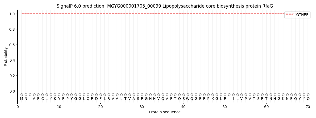
| Other | SP_Sec_SPI | LIPO_Sec_SPII | TAT_Tat_SPI | TATLIP_Sec_SPII | PILIN_Sec_SPIII |
|---|---|---|---|---|---|
| 1.000080 | 0.000000 | 0.000000 | 0.000000 | 0.000000 | 0.000000 |