You are browsing environment: HUMAN GUT
CAZyme Information: MGYG000001705_00105
You are here: Home > Sequence: MGYG000001705_00105
Basic Information |
Genomic context |
Full Sequence |
Enzyme annotations |
CAZy signature domains |
CDD domains |
CAZyme hits |
PDB hits |
Swiss-Prot hits |
SignalP and Lipop annotations |
TMHMM annotations
Basic Information help
| Species | Citrobacter portucalensis | |||||||||||
|---|---|---|---|---|---|---|---|---|---|---|---|---|
| Lineage | Bacteria; Proteobacteria; Gammaproteobacteria; Enterobacterales; Enterobacteriaceae; Citrobacter; Citrobacter portucalensis | |||||||||||
| CAZyme ID | MGYG000001705_00105 | |||||||||||
| CAZy Family | GT2 | |||||||||||
| CAZyme Description | Undecaprenyl-phosphate 4-deoxy-4-formamido-L-arabinose transferase | |||||||||||
| CAZyme Property |
|
|||||||||||
| Genome Property |
|
|||||||||||
| Gene Location | Start: 104763; End: 105776 Strand: - | |||||||||||
CAZyme Signature Domains help
| Family | Start | End | Evalue | family coverage |
|---|---|---|---|---|
| GT2 | 10 | 141 | 5.1e-28 | 0.7705882352941177 |
CDD Domains download full data without filtering help
| Cdd ID | Domain | E-Value | qStart | qEnd | sStart | sEnd | Domain Description |
|---|---|---|---|---|---|---|---|
| cd00761 | Glyco_tranf_GTA_type | 4.11e-28 | 11 | 121 | 1 | 111 | Glycosyltransferase family A (GT-A) includes diverse families of glycosyl transferases with a common GT-A type structural fold. Glycosyltransferases (GTs) are enzymes that synthesize oligosaccharides, polysaccharides, and glycoconjugates by transferring the sugar moiety from an activated nucleotide-sugar donor to an acceptor molecule, which may be a growing oligosaccharide, a lipid, or a protein. Based on the stereochemistry of the donor and acceptor molecules, GTs are classified as either retaining or inverting enzymes. To date, all GT structures adopt one of two possible folds, termed GT-A fold and GT-B fold. This hierarchy includes diverse families of glycosyl transferases with a common GT-A type structural fold, which has two tightly associated beta/alpha/beta domains that tend to form a continuous central sheet of at least eight beta-strands. The majority of the proteins in this superfamily are Glycosyltransferase family 2 (GT-2) proteins. But it also includes families GT-43, GT-6, GT-8, GT13 and GT-7; which are evolutionarily related to GT-2 and share structure similarities. |
| pfam00535 | Glycos_transf_2 | 7.11e-26 | 10 | 121 | 1 | 111 | Glycosyl transferase family 2. Diverse family, transferring sugar from UDP-glucose, UDP-N-acetyl- galactosamine, GDP-mannose or CDP-abequose, to a range of substrates including cellulose, dolichol phosphate and teichoic acids. |
| PRK10073 | PRK10073 | 3.75e-25 | 8 | 127 | 7 | 124 | putative glycosyl transferase; Provisional |
| COG0463 | WcaA | 1.18e-19 | 8 | 286 | 4 | 274 | Glycosyltransferase involved in cell wall bisynthesis [Cell wall/membrane/envelope biogenesis]. |
| cd06423 | CESA_like | 2.01e-19 | 11 | 121 | 1 | 112 | CESA_like is the cellulose synthase superfamily. The cellulose synthase (CESA) superfamily includes a wide variety of glycosyltransferase family 2 enzymes that share the common characteristic of catalyzing the elongation of polysaccharide chains. The members include cellulose synthase catalytic subunit, chitin synthase, glucan biosynthesis protein and other families of CESA-like proteins. Cellulose synthase catalyzes the polymerization reaction of cellulose, an aggregate of unbranched polymers of beta-1,4-linked glucose residues in plants, most algae, some bacteria and fungi, and even some animals. In bacteria, algae and lower eukaryotes, there is a second unrelated type of cellulose synthase (Type II), which produces acylated cellulose, a derivative of cellulose. Chitin synthase catalyzes the incorporation of GlcNAc from substrate UDP-GlcNAc into chitin, which is a linear homopolymer of beta-(1,4)-linked GlcNAc residues and Glucan Biosynthesis protein catalyzes the elongation of beta-1,2 polyglucose chains of Glucan. |
CAZyme Hits help
| Hit ID | E-Value | Query Start | Query End | Hit Start | Hit End |
|---|---|---|---|---|---|
| QNM34015.1 | 2.03e-248 | 1 | 337 | 1 | 337 |
| QNM28782.1 | 2.03e-248 | 1 | 337 | 1 | 337 |
| QNM24092.1 | 2.03e-248 | 1 | 337 | 1 | 337 |
| QNM18629.1 | 2.03e-248 | 1 | 337 | 1 | 337 |
| AUV41562.1 | 2.27e-245 | 1 | 337 | 1 | 337 |
PDB Hits download full data without filtering help
| Hit ID | E-Value | Query Start | Query End | Hit Start | Hit End | Description |
|---|---|---|---|---|---|---|
| 5HEA_A | 8.97e-16 | 5 | 235 | 3 | 234 | CgTstructure in hexamer [Streptococcus parasanguinis FW213],5HEA_B CgT structure in hexamer [Streptococcus parasanguinis FW213],5HEA_C CgT structure in hexamer [Streptococcus parasanguinis FW213],5HEC_A CgT structure in dimer [Streptococcus parasanguinis FW213],5HEC_B CgT structure in dimer [Streptococcus parasanguinis FW213] |
| 3L7I_A | 5.04e-11 | 9 | 115 | 4 | 110 | Structureof the Wall Teichoic Acid Polymerase TagF [Staphylococcus epidermidis RP62A],3L7I_B Structure of the Wall Teichoic Acid Polymerase TagF [Staphylococcus epidermidis RP62A],3L7I_C Structure of the Wall Teichoic Acid Polymerase TagF [Staphylococcus epidermidis RP62A],3L7I_D Structure of the Wall Teichoic Acid Polymerase TagF [Staphylococcus epidermidis RP62A] |
| 3L7J_A | 5.04e-11 | 9 | 115 | 4 | 110 | ChainA, Teichoic acid biosynthesis protein F [Staphylococcus epidermidis RP62A],3L7J_B Chain B, Teichoic acid biosynthesis protein F [Staphylococcus epidermidis RP62A],3L7J_C Chain C, Teichoic acid biosynthesis protein F [Staphylococcus epidermidis RP62A],3L7J_D Chain D, Teichoic acid biosynthesis protein F [Staphylococcus epidermidis RP62A],3L7K_A Chain A, Teichoic acid biosynthesis protein F [Staphylococcus epidermidis RP62A],3L7K_B Chain B, Teichoic acid biosynthesis protein F [Staphylococcus epidermidis RP62A],3L7K_C Chain C, Teichoic acid biosynthesis protein F [Staphylococcus epidermidis RP62A],3L7K_D Chain D, Teichoic acid biosynthesis protein F [Staphylococcus epidermidis RP62A],3L7L_A Chain A, Teichoic acid biosynthesis protein F [Staphylococcus epidermidis RP62A],3L7L_B Chain B, Teichoic acid biosynthesis protein F [Staphylococcus epidermidis RP62A],3L7L_C Chain C, Teichoic acid biosynthesis protein F [Staphylococcus epidermidis RP62A],3L7L_D Chain D, Teichoic acid biosynthesis protein F [Staphylococcus epidermidis RP62A] |
| 3L7M_A | 5.04e-11 | 9 | 115 | 4 | 110 | ChainA, Teichoic acid biosynthesis protein F [Staphylococcus epidermidis RP62A],3L7M_B Chain B, Teichoic acid biosynthesis protein F [Staphylococcus epidermidis RP62A],3L7M_C Chain C, Teichoic acid biosynthesis protein F [Staphylococcus epidermidis RP62A],3L7M_D Chain D, Teichoic acid biosynthesis protein F [Staphylococcus epidermidis RP62A] |
| 2Z87_A | 1.11e-09 | 8 | 116 | 375 | 481 | Crystalstructure of chondroitin polymerase from Escherichia coli strain K4 (K4CP) complexed with UDP-GalNAc and UDP [Escherichia coli],2Z87_B Crystal structure of chondroitin polymerase from Escherichia coli strain K4 (K4CP) complexed with UDP-GalNAc and UDP [Escherichia coli] |
Swiss-Prot Hits download full data without filtering help
| Hit ID | E-Value | Query Start | Query End | Hit Start | Hit End | Description |
|---|---|---|---|---|---|---|
| P71057 | 5.00e-16 | 10 | 262 | 7 | 261 | Putative glycosyltransferase EpsH OS=Bacillus subtilis (strain 168) OX=224308 GN=epsH PE=2 SV=1 |
| A0A0H2UR96 | 2.06e-15 | 8 | 229 | 4 | 227 | Glycosyltransferase GlyG OS=Streptococcus pneumoniae serotype 4 (strain ATCC BAA-334 / TIGR4) OX=170187 GN=glyG PE=1 SV=1 |
| P71059 | 5.73e-15 | 8 | 225 | 4 | 220 | Uncharacterized glycosyltransferase EpsJ OS=Bacillus subtilis (strain 168) OX=224308 GN=epsJ PE=2 SV=1 |
| A0A0H2URH7 | 7.68e-15 | 9 | 120 | 7 | 116 | Glycosyltransferase GlyA OS=Streptococcus pneumoniae serotype 4 (strain ATCC BAA-334 / TIGR4) OX=170187 GN=glyA PE=3 SV=1 |
| P11290 | 1.18e-13 | 1 | 124 | 1 | 121 | Uncharacterized glycosyltransferase YibD OS=Escherichia coli (strain K12) OX=83333 GN=yibD PE=3 SV=2 |
SignalP and Lipop Annotations help
This protein is predicted as OTHER
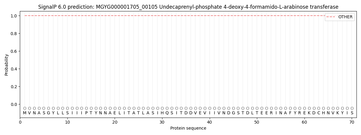
| Other | SP_Sec_SPI | LIPO_Sec_SPII | TAT_Tat_SPI | TATLIP_Sec_SPII | PILIN_Sec_SPIII |
|---|---|---|---|---|---|
| 1.000054 | 0.000000 | 0.000000 | 0.000000 | 0.000000 | 0.000000 |