You are browsing environment: HUMAN GUT
CAZyme Information: MGYG000001805_00379
You are here: Home > Sequence: MGYG000001805_00379
Basic Information |
Genomic context |
Full Sequence |
Enzyme annotations |
CAZy signature domains |
CDD domains |
CAZyme hits |
PDB hits |
Swiss-Prot hits |
SignalP and Lipop annotations |
TMHMM annotations
Basic Information help
| Species | ||||||||||||
|---|---|---|---|---|---|---|---|---|---|---|---|---|
| Lineage | Bacteria; Bacteroidota; Bacteroidia; Bacteroidales; Bacteroidaceae; Prevotella; | |||||||||||
| CAZyme ID | MGYG000001805_00379 | |||||||||||
| CAZy Family | GT2 | |||||||||||
| CAZyme Description | 2,3,4,5-tetrahydropyridine-2,6-dicarboxylate N-acetyltransferase | |||||||||||
| CAZyme Property |
|
|||||||||||
| Genome Property |
|
|||||||||||
| Gene Location | Start: 5303; End: 6568 Strand: + | |||||||||||
CAZyme Signature Domains help
| Family | Start | End | Evalue | family coverage |
|---|---|---|---|---|
| GT2 | 6 | 125 | 2.4e-21 | 0.7176470588235294 |
CDD Domains download full data without filtering help
| Cdd ID | Domain | E-Value | qStart | qEnd | sStart | sEnd | Domain Description |
|---|---|---|---|---|---|---|---|
| cd06433 | GT_2_WfgS_like | 6.36e-60 | 6 | 206 | 1 | 201 | WfgS and WfeV are involved in O-antigen biosynthesis. Escherichia coli WfgS and Shigella dysenteriae WfeV are glycosyltransferase 2 family enzymes involved in O-antigen biosynthesis. GT-2 enzymes have GT-A type structural fold, which has two tightly associated beta/alpha/beta domains that tend to form a continuous central sheet of at least eight beta-strands. These are enzymes that catalyze the transfer of sugar moieties from activated donor molecules to specific acceptor molecules, forming glycosidic bonds. Glycosyltransferases have been classified into more than 90 distinct sequence based families. |
| cd04647 | LbH_MAT_like | 8.68e-37 | 300 | 403 | 2 | 109 | Maltose O-acyltransferase (MAT)-like: This family is composed of maltose O-acetyltransferase, galactoside O-acetyltransferase (GAT), xenobiotic acyltransferase (XAT) and similar proteins. MAT and GAT catalyze the CoA-dependent acetylation of the 6-hydroxyl group of their respective sugar substrates. MAT acetylates maltose and glucose exclusively while GAT specifically acetylates galactopyranosides. XAT catalyzes the CoA-dependent acetylation of a variety of hydroxyl-bearing acceptors such as chloramphenicol and streptogramin, among others. XATs are implicated in inactivating xenobiotics leading to xenobiotic resistance in patients. Members of this family contain a a left-handed parallel beta-helix (LbH) domain with at least 5 turns, each containing three imperfect tandem repeats of a hexapeptide repeat motif (X-[STAV]-X-[LIV]-[GAED]-X). They are trimeric in their active form. |
| PRK10502 | PRK10502 | 3.29e-28 | 256 | 407 | 25 | 179 | putative acyl transferase; Provisional |
| cd05825 | LbH_wcaF_like | 5.16e-28 | 300 | 403 | 4 | 107 | wcaF-like: This group is composed of the protein product of the E. coli wcaF gene and similar proteins. WcaF is part of the gene cluster responsible for the biosynthesis of the extracellular polysaccharide colanic acid. The wcaF protein is predicted to contain a left-handed parallel beta-helix (LbH) domain encoded by imperfect tandem repeats of a hexapeptide repeat motif (X-[STAV]-X-[LIV]-[GAED]-X). Proteins containing hexapeptide repeats are often enzymes showing acyltransferase activity. Many are trimeric in their active forms. |
| cd03357 | LbH_MAT_GAT | 2.69e-23 | 271 | 403 | 34 | 169 | Maltose O-acetyltransferase (MAT) and Galactoside O-acetyltransferase (GAT): MAT and GAT catalyze the CoA-dependent acetylation of the 6-hydroxyl group of their respective sugar substrates. MAT acetylates maltose and glucose exclusively at the C6 position of the nonreducing end glucosyl moiety. GAT specifically acetylates galactopyranosides. Furthermore, MAT shows higher affinity toward artificial substrates containing an alkyl or hydrophobic chain as well as a glucosyl unit. Active MAT and GAT are homotrimers, with each subunit consisting of an N-terminal alpha-helical region and a C-terminal left-handed parallel alpha-helix (LbH) subdomain with 6 turns, each containing three imperfect tandem repeats of a hexapeptide repeat motif (X-[STAV]-X-[LIV]-[GAED]-X). |
CAZyme Hits help
| Hit ID | E-Value | Query Start | Query End | Hit Start | Hit End |
|---|---|---|---|---|---|
| QUI93290.1 | 1.44e-169 | 1 | 421 | 1 | 421 |
| AXV50072.1 | 5.81e-169 | 1 | 421 | 1 | 421 |
| QUB91771.1 | 1.17e-168 | 1 | 421 | 1 | 421 |
| QJR60740.1 | 7.25e-58 | 1 | 246 | 1 | 243 |
| QJR54460.1 | 7.25e-58 | 1 | 246 | 1 | 243 |
PDB Hits download full data without filtering help
| Hit ID | E-Value | Query Start | Query End | Hit Start | Hit End | Description |
|---|---|---|---|---|---|---|
| 3VBP_A | 1.53e-08 | 275 | 410 | 27 | 192 | CrystalStructure of the D94N mutant of AntD, an N-acyltransferase from Bacillus cereus in complex with dTDP and Coenzyme A [Bacillus cereus SJ1],3VBP_C Crystal Structure of the D94N mutant of AntD, an N-acyltransferase from Bacillus cereus in complex with dTDP and Coenzyme A [Bacillus cereus SJ1],3VBP_E Crystal Structure of the D94N mutant of AntD, an N-acyltransferase from Bacillus cereus in complex with dTDP and Coenzyme A [Bacillus cereus SJ1] |
| 3VBK_A | 2.80e-08 | 275 | 410 | 27 | 192 | CrystalStructure of the S84A mutant of AntD, an N-acyltransferase from Bacillus cereus in complex with dTDP-4-amino-4,6-dideoxyglucose and Coenzyme A [Bacillus cereus SJ1],3VBK_C Crystal Structure of the S84A mutant of AntD, an N-acyltransferase from Bacillus cereus in complex with dTDP-4-amino-4,6-dideoxyglucose and Coenzyme A [Bacillus cereus SJ1],3VBK_E Crystal Structure of the S84A mutant of AntD, an N-acyltransferase from Bacillus cereus in complex with dTDP-4-amino-4,6-dideoxyglucose and Coenzyme A [Bacillus cereus SJ1] |
| 3VBI_A | 2.80e-08 | 275 | 410 | 27 | 192 | CrystalStructure of AntD, an N-acyltransferase from Bacillus cereus in complex with dTDP-4-amino-4,6-dideoxyglucose and Coenzyme A [Bacillus cereus SJ1],3VBI_C Crystal Structure of AntD, an N-acyltransferase from Bacillus cereus in complex with dTDP-4-amino-4,6-dideoxyglucose and Coenzyme A [Bacillus cereus SJ1],3VBI_E Crystal Structure of AntD, an N-acyltransferase from Bacillus cereus in complex with dTDP-4-amino-4,6-dideoxyglucose and Coenzyme A [Bacillus cereus SJ1],3VBJ_A Crystal Structure of AntD, an N-acyltransferase from Bacillus cereus in complex with dTDP and 3-hydroxybutyryl-CoA [Bacillus cereus SJ1],3VBJ_C Crystal Structure of AntD, an N-acyltransferase from Bacillus cereus in complex with dTDP and 3-hydroxybutyryl-CoA [Bacillus cereus SJ1],3VBJ_E Crystal Structure of AntD, an N-acyltransferase from Bacillus cereus in complex with dTDP and 3-hydroxybutyryl-CoA [Bacillus cereus SJ1] |
| 3VBL_A | 2.80e-08 | 275 | 410 | 27 | 192 | CrystalStructure of the S84C mutant of AntD, an N-acyltransferase from Bacillus cereus in complex with dTDP-4-amino-4,6-dideoxyglucose and Coenzyme A [Bacillus cereus SJ1],3VBL_C Crystal Structure of the S84C mutant of AntD, an N-acyltransferase from Bacillus cereus in complex with dTDP-4-amino-4,6-dideoxyglucose and Coenzyme A [Bacillus cereus SJ1],3VBL_E Crystal Structure of the S84C mutant of AntD, an N-acyltransferase from Bacillus cereus in complex with dTDP-4-amino-4,6-dideoxyglucose and Coenzyme A [Bacillus cereus SJ1] |
| 3VBM_A | 2.80e-08 | 275 | 410 | 27 | 192 | CrystalStructure of the S84T mutant of AntD, an N-acyltransferase from Bacillus cereus in complex with dTDP and Coenzyme A [Bacillus cereus SJ1],3VBM_C Crystal Structure of the S84T mutant of AntD, an N-acyltransferase from Bacillus cereus in complex with dTDP and Coenzyme A [Bacillus cereus SJ1],3VBM_E Crystal Structure of the S84T mutant of AntD, an N-acyltransferase from Bacillus cereus in complex with dTDP and Coenzyme A [Bacillus cereus SJ1] |
Swiss-Prot Hits download full data without filtering help
| Hit ID | E-Value | Query Start | Query End | Hit Start | Hit End | Description |
|---|---|---|---|---|---|---|
| P71239 | 1.48e-15 | 5 | 204 | 3 | 201 | Putative colanic acid biosynthesis glycosyl transferase WcaE OS=Escherichia coli (strain K12) OX=83333 GN=wcaE PE=4 SV=2 |
| P9WMX9 | 6.23e-15 | 5 | 93 | 7 | 96 | Uncharacterized glycosyltransferase Rv1514c OS=Mycobacterium tuberculosis (strain ATCC 25618 / H37Rv) OX=83332 GN=Rv1514c PE=1 SV=1 |
| P9WMX8 | 6.23e-15 | 5 | 93 | 7 | 96 | Uncharacterized glycosyltransferase MT1564 OS=Mycobacterium tuberculosis (strain CDC 1551 / Oshkosh) OX=83331 GN=MT1564 PE=3 SV=1 |
| A0A0H2UR96 | 3.22e-11 | 1 | 123 | 1 | 125 | Glycosyltransferase GlyG OS=Streptococcus pneumoniae serotype 4 (strain ATCC BAA-334 / TIGR4) OX=170187 GN=glyG PE=1 SV=1 |
| A0A0H2URH7 | 9.61e-11 | 1 | 123 | 3 | 126 | Glycosyltransferase GlyA OS=Streptococcus pneumoniae serotype 4 (strain ATCC BAA-334 / TIGR4) OX=170187 GN=glyA PE=3 SV=1 |
SignalP and Lipop Annotations help
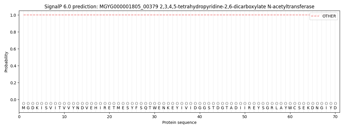
This protein is predicted as OTHER
| Other | SP_Sec_SPI | LIPO_Sec_SPII | TAT_Tat_SPI | TATLIP_Sec_SPII | PILIN_Sec_SPIII |
|---|---|---|---|---|---|
| 1.000075 | 0.000000 | 0.000000 | 0.000000 | 0.000000 | 0.000000 |