You are browsing environment: HUMAN GUT
CAZyme Information: MGYG000001878_00322
You are here: Home > Sequence: MGYG000001878_00322
Basic Information |
Genomic context |
Full Sequence |
Enzyme annotations |
CAZy signature domains |
CDD domains |
CAZyme hits |
PDB hits |
Swiss-Prot hits |
SignalP and Lipop annotations |
TMHMM annotations
Basic Information help
| Species | Parabacteroides sp900548175 | |||||||||||
|---|---|---|---|---|---|---|---|---|---|---|---|---|
| Lineage | Bacteria; Bacteroidota; Bacteroidia; Bacteroidales; Tannerellaceae; Parabacteroides; Parabacteroides sp900548175 | |||||||||||
| CAZyme ID | MGYG000001878_00322 | |||||||||||
| CAZy Family | GH109 | |||||||||||
| CAZyme Description | Inositol 2-dehydrogenase/D-chiro-inositol 3-dehydrogenase | |||||||||||
| CAZyme Property |
|
|||||||||||
| Genome Property |
|
|||||||||||
| Gene Location | Start: 28091; End: 29476 Strand: + | |||||||||||
CAZyme Signature Domains help
| Family | Start | End | Evalue | family coverage |
|---|---|---|---|---|
| GH109 | 69 | 257 | 5.4e-18 | 0.45864661654135336 |
CDD Domains download full data without filtering help
| Cdd ID | Domain | E-Value | qStart | qEnd | sStart | sEnd | Domain Description |
|---|---|---|---|---|---|---|---|
| COG0673 | MviM | 1.33e-36 | 68 | 419 | 1 | 335 | Predicted dehydrogenase [General function prediction only]. |
| pfam01408 | GFO_IDH_MocA | 2.17e-11 | 71 | 196 | 1 | 115 | Oxidoreductase family, NAD-binding Rossmann fold. This family of enzymes utilize NADP or NAD. This family is called the GFO/IDH/MOCA family in swiss-prot. |
| pfam03435 | Sacchrp_dh_NADP | 7.29e-06 | 75 | 201 | 3 | 120 | Saccharopine dehydrogenase NADP binding domain. This family contains the NADP binding domain of saccharopine dehydrogenase. In some organisms this enzyme is found as a bifunctional polypeptide with lysine ketoglutarate reductase. The saccharopine dehydrogenase can also function as a saccharopine reductase. |
| pfam10518 | TAT_signal | 1.81e-05 | 7 | 31 | 2 | 26 | TAT (twin-arginine translocation) pathway signal sequence. |
| TIGR01299 | synapt_SV2 | 0.002 | 282 | 320 | 467 | 506 | synaptic vesicle protein SV2. This model describes a tightly conserved subfamily of the larger family of sugar (and other) transporters described by pfam00083. Members of this subfamily include closely related forms SV2A and SV2B of synaptic vesicle protein from vertebrates and a more distantly related homolog (below trusted cutoff) from Drosophila melanogaster. Members are predicted to have two sets of six transmembrane helices. |
CAZyme Hits help
| Hit ID | E-Value | Query Start | Query End | Hit Start | Hit End |
|---|---|---|---|---|---|
| BBK93797.1 | 2.02e-306 | 1 | 461 | 1 | 460 |
| QIX65768.1 | 2.02e-306 | 1 | 461 | 1 | 460 |
| QUT96603.1 | 2.02e-306 | 1 | 461 | 1 | 460 |
| AST52349.1 | 2.02e-306 | 1 | 461 | 1 | 460 |
| QUT20197.1 | 2.02e-306 | 1 | 461 | 1 | 460 |
PDB Hits download full data without filtering help
| Hit ID | E-Value | Query Start | Query End | Hit Start | Hit End | Description |
|---|---|---|---|---|---|---|
| 3CEA_A | 7.48e-09 | 66 | 296 | 4 | 210 | ChainA, Myo-inositol 2-dehydrogenase [Lactiplantibacillus plantarum WCFS1],3CEA_B Chain B, Myo-inositol 2-dehydrogenase [Lactiplantibacillus plantarum WCFS1],3CEA_C Chain C, Myo-inositol 2-dehydrogenase [Lactiplantibacillus plantarum WCFS1],3CEA_D Chain D, Myo-inositol 2-dehydrogenase [Lactiplantibacillus plantarum WCFS1] |
| 3EC7_A | 4.26e-07 | 68 | 226 | 21 | 168 | CrystalStructure of Putative Dehydrogenase from Salmonella typhimurium LT2 [Salmonella enterica subsp. enterica serovar Typhimurium str. LT2],3EC7_B Crystal Structure of Putative Dehydrogenase from Salmonella typhimurium LT2 [Salmonella enterica subsp. enterica serovar Typhimurium str. LT2],3EC7_C Crystal Structure of Putative Dehydrogenase from Salmonella typhimurium LT2 [Salmonella enterica subsp. enterica serovar Typhimurium str. LT2],3EC7_D Crystal Structure of Putative Dehydrogenase from Salmonella typhimurium LT2 [Salmonella enterica subsp. enterica serovar Typhimurium str. LT2],3EC7_E Crystal Structure of Putative Dehydrogenase from Salmonella typhimurium LT2 [Salmonella enterica subsp. enterica serovar Typhimurium str. LT2],3EC7_F Crystal Structure of Putative Dehydrogenase from Salmonella typhimurium LT2 [Salmonella enterica subsp. enterica serovar Typhimurium str. LT2],3EC7_G Crystal Structure of Putative Dehydrogenase from Salmonella typhimurium LT2 [Salmonella enterica subsp. enterica serovar Typhimurium str. LT2],3EC7_H Crystal Structure of Putative Dehydrogenase from Salmonella typhimurium LT2 [Salmonella enterica subsp. enterica serovar Typhimurium str. LT2] |
Swiss-Prot Hits download full data without filtering help
| Hit ID | E-Value | Query Start | Query End | Hit Start | Hit End | Description |
|---|---|---|---|---|---|---|
| Q9RK81 | 4.66e-09 | 1 | 230 | 1 | 213 | Glycosyl hydrolase family 109 protein OS=Streptomyces coelicolor (strain ATCC BAA-471 / A3(2) / M145) OX=100226 GN=SCO0529 PE=3 SV=1 |
| A9N564 | 1.62e-06 | 71 | 321 | 3 | 222 | Inositol 2-dehydrogenase OS=Salmonella paratyphi B (strain ATCC BAA-1250 / SPB7) OX=1016998 GN=iolG PE=3 SV=1 |
| B5F3F4 | 1.62e-06 | 71 | 321 | 3 | 222 | Inositol 2-dehydrogenase OS=Salmonella agona (strain SL483) OX=454166 GN=iolG PE=3 SV=1 |
| Q8ZK57 | 1.62e-06 | 71 | 321 | 3 | 222 | Inositol 2-dehydrogenase OS=Salmonella typhimurium (strain LT2 / SGSC1412 / ATCC 700720) OX=99287 GN=iolG PE=1 SV=1 |
SignalP and Lipop Annotations help
This protein is predicted as TAT
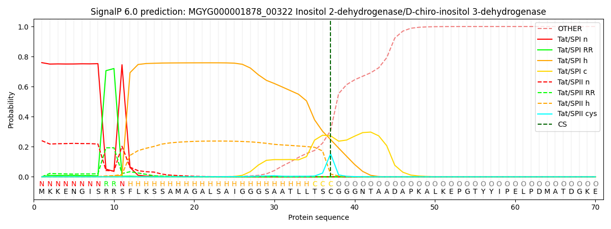
| Other | SP_Sec_SPI | LIPO_Sec_SPII | TAT_Tat_SPI | TATLIP_Sec_SPII | PILIN_Sec_SPIII |
|---|---|---|---|---|---|
| 0.000001 | 0.000000 | 0.000004 | 0.759618 | 0.240380 | 0.000000 |