You are browsing environment: HUMAN GUT
CAZyme Information: MGYG000001884_02014
You are here: Home > Sequence: MGYG000001884_02014
Basic Information |
Genomic context |
Full Sequence |
Enzyme annotations |
CAZy signature domains |
CDD domains |
CAZyme hits |
PDB hits |
Swiss-Prot hits |
SignalP and Lipop annotations |
TMHMM annotations
Basic Information help
| Species | UBA1829 sp900760615 | |||||||||||
|---|---|---|---|---|---|---|---|---|---|---|---|---|
| Lineage | Bacteria; Verrucomicrobiota; Lentisphaeria; Victivallales; UBA1829; UBA1829; UBA1829 sp900760615 | |||||||||||
| CAZyme ID | MGYG000001884_02014 | |||||||||||
| CAZy Family | GH38 | |||||||||||
| CAZyme Description | hypothetical protein | |||||||||||
| CAZyme Property |
|
|||||||||||
| Genome Property |
|
|||||||||||
| Gene Location | Start: 172; End: 1650 Strand: + | |||||||||||
CDD Domains download full data without filtering help
| Cdd ID | Domain | E-Value | qStart | qEnd | sStart | sEnd | Domain Description |
|---|---|---|---|---|---|---|---|
| COG0383 | AMS1 | 1.12e-71 | 17 | 490 | 491 | 941 | Alpha-mannosidase [Carbohydrate transport and metabolism]. |
| pfam07748 | Glyco_hydro_38C | 1.01e-62 | 155 | 358 | 1 | 202 | Glycosyl hydrolases family 38 C-terminal domain. Glycosyl hydrolases are key enzymes of carbohydrate metabolism. |
| pfam09261 | Alpha-mann_mid | 3.36e-18 | 1 | 72 | 23 | 98 | Alpha mannosidase middle domain. Members of this family adopt a structure consisting of three alpha helices, in an immunoglobulin/albumin-binding domain-like fold. They are predominantly found in the enzyme alpha-mannosidase. |
| pfam17677 | Glyco_hydro38C2 | 1.14e-16 | 415 | 489 | 1 | 73 | Glycosyl hydrolases family 38 C-terminal beta sandwich domain. This domain is found at the C-terminal end of various glycosyl hydrolases belonging to family 38. The domain has a beta sandwich fold. |
| smart00872 | Alpha-mann_mid | 5.23e-16 | 1 | 53 | 22 | 78 | Alpha mannosidase, middle domain. Members of this entry belong to the glycosyl hydrolase family 38, This domain, which is found in the central region adopts a structure consisting of three alpha helices, in an immunoglobulin/albumin-binding domain-like fold. The domain is predominantly found in the enzyme alpha-mannosidase. |
CAZyme Hits help
| Hit ID | E-Value | Query Start | Query End | Hit Start | Hit End |
|---|---|---|---|---|---|
| AVM46843.1 | 4.07e-159 | 1 | 491 | 529 | 1017 |
| AVM45569.1 | 8.12e-153 | 1 | 490 | 534 | 1021 |
| AVM43446.1 | 5.28e-151 | 1 | 491 | 539 | 1025 |
| APF18337.1 | 3.39e-135 | 2 | 490 | 545 | 1034 |
| CCW34438.1 | 2.00e-96 | 6 | 490 | 535 | 1055 |
PDB Hits download full data without filtering help
| Hit ID | E-Value | Query Start | Query End | Hit Start | Hit End | Description |
|---|---|---|---|---|---|---|
| 6LZ1_A | 2.41e-55 | 17 | 490 | 597 | 1075 | Structureof S.pombe alpha-mannosidase Ams1 [Schizosaccharomyces pombe 972h-],6LZ1_B Structure of S.pombe alpha-mannosidase Ams1 [Schizosaccharomyces pombe 972h-],6LZ1_C Structure of S.pombe alpha-mannosidase Ams1 [Schizosaccharomyces pombe 972h-],6LZ1_D Structure of S.pombe alpha-mannosidase Ams1 [Schizosaccharomyces pombe 972h-] |
| 7DD9_A | 2.97e-55 | 17 | 490 | 597 | 1075 | ChainA, Alpha-mannosidase,ZZ-type zinc finger-containing protein P35G2.11c,Maltose/maltodextrin-binding periplasmic protein [synthetic construct],7DD9_C Chain C, Alpha-mannosidase,ZZ-type zinc finger-containing protein P35G2.11c,Maltose/maltodextrin-binding periplasmic protein [synthetic construct],7DD9_E Chain E, Alpha-mannosidase,ZZ-type zinc finger-containing protein P35G2.11c,Maltose/maltodextrin-binding periplasmic protein [synthetic construct],7DD9_G Chain G, Alpha-mannosidase,ZZ-type zinc finger-containing protein P35G2.11c,Maltose/maltodextrin-binding periplasmic protein [synthetic construct] |
| 5JM0_A | 1.06e-49 | 16 | 490 | 620 | 1094 | Structureof the S. cerevisiae alpha-mannosidase 1 [Saccharomyces cerevisiae S288C] |
| 2WYH_A | 1.08e-09 | 17 | 478 | 355 | 915 | Structureof the Streptococcus pyogenes family GH38 alpha-mannosidase [Streptococcus pyogenes M1 GAS],2WYH_B Structure of the Streptococcus pyogenes family GH38 alpha-mannosidase [Streptococcus pyogenes M1 GAS],2WYI_A Structure of the Streptococcus pyogenes family GH38 alpha-mannosidase complexed with swainsonine [Streptococcus pyogenes M1 GAS],2WYI_B Structure of the Streptococcus pyogenes family GH38 alpha-mannosidase complexed with swainsonine [Streptococcus pyogenes M1 GAS] |
| 5KBP_A | 1.19e-07 | 17 | 491 | 335 | 898 | Thecrystal structure of an alpha-mannosidase from Enterococcus faecalis V583 [Enterococcus faecalis V583],5KBP_B The crystal structure of an alpha-mannosidase from Enterococcus faecalis V583 [Enterococcus faecalis V583] |
Swiss-Prot Hits download full data without filtering help
| Hit ID | E-Value | Query Start | Query End | Hit Start | Hit End | Description |
|---|---|---|---|---|---|---|
| Q9NTJ4 | 5.32e-75 | 17 | 490 | 559 | 1033 | Alpha-mannosidase 2C1 OS=Homo sapiens OX=9606 GN=MAN2C1 PE=1 SV=1 |
| Q91W89 | 3.61e-74 | 17 | 490 | 558 | 1032 | Alpha-mannosidase 2C1 OS=Mus musculus OX=10090 GN=Man2c1 PE=1 SV=1 |
| P21139 | 1.95e-66 | 17 | 490 | 558 | 1033 | Alpha-mannosidase 2C1 OS=Rattus norvegicus OX=10116 GN=Man2c1 PE=1 SV=1 |
| Q54K67 | 2.03e-64 | 5 | 489 | 547 | 1083 | Alpha-mannosidase G OS=Dictyostelium discoideum OX=44689 GN=manG PE=1 SV=1 |
| Q9UT61 | 1.20e-54 | 17 | 490 | 597 | 1075 | Alpha-mannosidase OS=Schizosaccharomyces pombe (strain 972 / ATCC 24843) OX=284812 GN=ams1 PE=1 SV=1 |
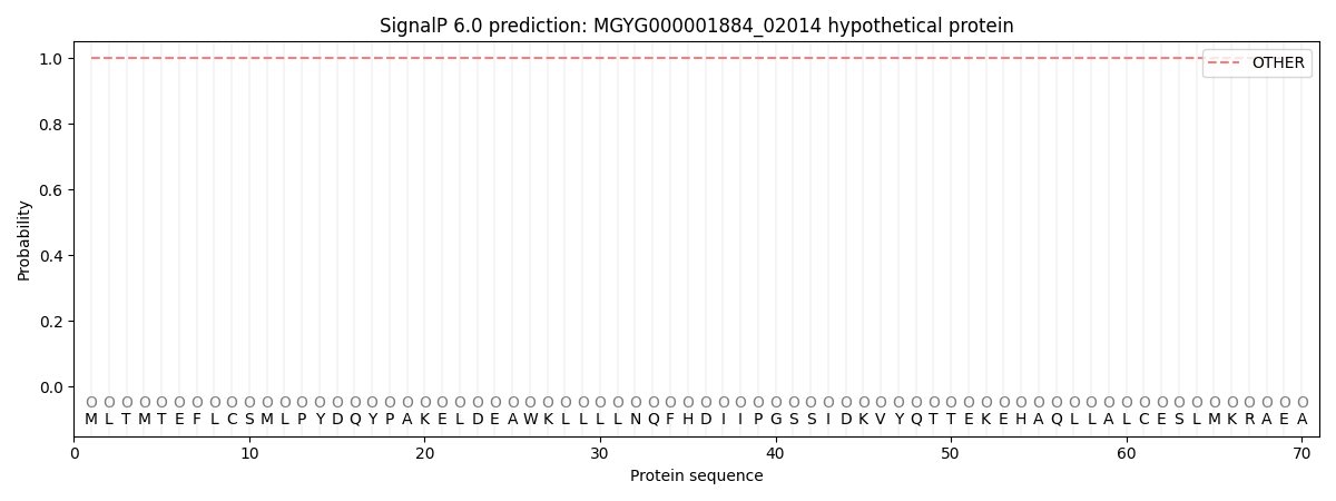
SignalP and Lipop Annotations help
This protein is predicted as OTHER
| Other | SP_Sec_SPI | LIPO_Sec_SPII | TAT_Tat_SPI | TATLIP_Sec_SPII | PILIN_Sec_SPIII |
|---|---|---|---|---|---|
| 1.000082 | 0.000000 | 0.000000 | 0.000000 | 0.000000 | 0.000000 |