You are browsing environment: HUMAN GUT
CAZyme Information: MGYG000001952_00004
You are here: Home > Sequence: MGYG000001952_00004
Basic Information |
Genomic context |
Full Sequence |
Enzyme annotations |
CAZy signature domains |
CDD domains |
CAZyme hits |
PDB hits |
Swiss-Prot hits |
SignalP and Lipop annotations |
TMHMM annotations
Basic Information help
| Species | ||||||||||||
|---|---|---|---|---|---|---|---|---|---|---|---|---|
| Lineage | Bacteria; Firmicutes_A; Clostridia; Lachnospirales; Lachnospiraceae; Lachnoclostridium_A; | |||||||||||
| CAZyme ID | MGYG000001952_00004 | |||||||||||
| CAZy Family | CBM50 | |||||||||||
| CAZyme Description | hypothetical protein | |||||||||||
| CAZyme Property |
|
|||||||||||
| Genome Property |
|
|||||||||||
| Gene Location | Start: 1998; End: 2657 Strand: - | |||||||||||
CDD Domains download full data without filtering help
| Cdd ID | Domain | E-Value | qStart | qEnd | sStart | sEnd | Domain Description |
|---|---|---|---|---|---|---|---|
| PRK11198 | PRK11198 | 8.78e-23 | 159 | 219 | 86 | 147 | LysM domain/BON superfamily protein; Provisional |
| COG1652 | XkdP | 4.93e-15 | 135 | 219 | 176 | 263 | Nucleoid-associated protein YgaU, contains BON and LysM domains [Function unknown]. |
| cd00118 | LysM | 1.36e-13 | 169 | 217 | 1 | 45 | Lysin Motif is a small domain involved in binding peptidoglycan. LysM, a small globular domain with approximately 40 amino acids, is a widespread protein module involved in binding peptidoglycan in bacteria and chitin in eukaryotes. The domain was originally identified in enzymes that degrade bacterial cell walls, but proteins involved in many other biological functions also contain this domain. It has been reported that the LysM domain functions as a signal for specific plant-bacteria recognition in bacterial pathogenesis. Many of these enzymes are modular and are composed of catalytic units linked to one or several repeats of LysM domains. LysM domains are found in bacteria and eukaryotes. |
| TIGR02899 | spore_safA | 5.90e-13 | 173 | 219 | 1 | 44 | spore coat assembly protein SafA. SafA (YrbB) (SafA) of Bacillus subtilis is a protein found at the interface of the spore cortex and spore coat, and is dependent on SpoVID for its localization. This model is based on the N-terminal LysM (lysin motif) domain (see pfamAM model pfam01476) of SafA and, from several other spore-forming species, the protein with the most similar N-terminal region. However, this set of proteins differs greatly in C-terminal of the LysM domaim; blocks of 12-residue and 13-residue repeats are found in the Bacillus cereus group, tandem LysM domains in Thermoanaerobacter tengcongensis MB4, etc. in which one of which is found in most examples of endospore-forming bacteria. [Cellular processes, Sporulation and germination] |
| pfam01476 | LysM | 1.30e-11 | 171 | 218 | 1 | 43 | LysM domain. The LysM (lysin motif) domain is about 40 residues long. It is found in a variety of enzymes involved in bacterial cell wall degradation. This domain may have a general peptidoglycan binding function. The structure of this domain is known. |
CAZyme Hits help
| Hit ID | E-Value | Query Start | Query End | Hit Start | Hit End |
|---|---|---|---|---|---|
| QRV21693.1 | 9.61e-106 | 1 | 219 | 1 | 220 |
| ADL04006.1 | 9.61e-106 | 1 | 219 | 1 | 220 |
| AGC67443.1 | 8.07e-59 | 1 | 217 | 1 | 224 |
| AGI38503.1 | 8.07e-59 | 1 | 217 | 1 | 224 |
| ABN53691.1 | 1.09e-58 | 1 | 218 | 1 | 218 |
PDB Hits download full data without filtering help
| Hit ID | E-Value | Query Start | Query End | Hit Start | Hit End | Description |
|---|---|---|---|---|---|---|
| 5FIM_A | 1.89e-10 | 166 | 218 | 94 | 147 | Thestructure of Kbp.K from E. coli [Escherichia coli],7PVC_A Chain A, Potassium binding protein Kbp [Escherichia coli K-12] |
Swiss-Prot Hits download full data without filtering help
| Hit ID | E-Value | Query Start | Query End | Hit Start | Hit End | Description |
|---|---|---|---|---|---|---|
| P54335 | 6.24e-18 | 2 | 218 | 5 | 218 | Phage-like element PBSX protein XkdP OS=Bacillus subtilis (strain 168) OX=224308 GN=xkdP PE=4 SV=2 |
| P45932 | 8.86e-17 | 3 | 218 | 6 | 218 | Uncharacterized protein YqbP OS=Bacillus subtilis (strain 168) OX=224308 GN=yqbP PE=4 SV=1 |
| P0ADE6 | 8.92e-10 | 166 | 218 | 94 | 147 | Potassium binding protein Kbp OS=Escherichia coli (strain K12) OX=83333 GN=kbp PE=1 SV=2 |
| P0ADE7 | 8.92e-10 | 166 | 218 | 94 | 147 | Potassium binding protein Kbp OS=Escherichia coli O6:H1 (strain CFT073 / ATCC 700928 / UPEC) OX=199310 GN=kbp PE=3 SV=2 |
| Q9RVY3 | 2.29e-06 | 171 | 218 | 206 | 252 | Uncharacterized protein DR_0888 OS=Deinococcus radiodurans (strain ATCC 13939 / DSM 20539 / JCM 16871 / LMG 4051 / NBRC 15346 / NCIMB 9279 / R1 / VKM B-1422) OX=243230 GN=DR_0888 PE=1 SV=1 |
SignalP and Lipop Annotations help
This protein is predicted as OTHER
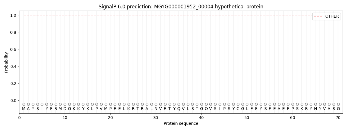
| Other | SP_Sec_SPI | LIPO_Sec_SPII | TAT_Tat_SPI | TATLIP_Sec_SPII | PILIN_Sec_SPIII |
|---|---|---|---|---|---|
| 1.000050 | 0.000000 | 0.000000 | 0.000000 | 0.000000 | 0.000000 |