You are browsing environment: HUMAN GUT
CAZyme Information: MGYG000002134_00001
You are here: Home > Sequence: MGYG000002134_00001
Basic Information |
Genomic context |
Full Sequence |
Enzyme annotations |
CAZy signature domains |
CDD domains |
CAZyme hits |
PDB hits |
Swiss-Prot hits |
SignalP and Lipop annotations |
TMHMM annotations
Basic Information help
| Species | Mailhella sp900541395 | |||||||||||
|---|---|---|---|---|---|---|---|---|---|---|---|---|
| Lineage | Bacteria; Desulfobacterota; Desulfovibrionia; Desulfovibrionales; Desulfovibrionaceae; Mailhella; Mailhella sp900541395 | |||||||||||
| CAZyme ID | MGYG000002134_00001 | |||||||||||
| CAZy Family | GT4 | |||||||||||
| CAZyme Description | D-inositol-3-phosphate glycosyltransferase | |||||||||||
| CAZyme Property |
|
|||||||||||
| Genome Property |
|
|||||||||||
| Gene Location | Start: 191; End: 1147 Strand: + | |||||||||||
CDD Domains download full data without filtering help
| Cdd ID | Domain | E-Value | qStart | qEnd | sStart | sEnd | Domain Description |
|---|---|---|---|---|---|---|---|
| cd03825 | GT4_WcaC-like | 9.17e-114 | 3 | 312 | 51 | 362 | putative colanic acid biosynthesis glycosyl transferase WcaC and similar proteins. This family is most closely related to the GT4 family of glycosyltransferases. Escherichia coli WcaC has been predicted to function in colanic acid biosynthesis. WcfI in Bacteroides fragilis has been shown to be involved in the capsular polysaccharide biosynthesis. |
| cd03801 | GT4_PimA-like | 6.24e-45 | 5 | 312 | 84 | 366 | phosphatidyl-myo-inositol mannosyltransferase. This family is most closely related to the GT4 family of glycosyltransferases and named after PimA in Propionibacterium freudenreichii, which is involved in the biosynthesis of phosphatidyl-myo-inositol mannosides (PIM) which are early precursors in the biosynthesis of lipomannans (LM) and lipoarabinomannans (LAM), and catalyzes the addition of a mannosyl residue from GDP-D-mannose (GDP-Man) to the position 2 of the carrier lipid phosphatidyl-myo-inositol (PI) to generate a phosphatidyl-myo-inositol bearing an alpha-1,2-linked mannose residue (PIM1). Glycosyltransferases catalyze the transfer of sugar moieties from activated donor molecules to specific acceptor molecules, forming glycosidic bonds. The acceptor molecule can be a lipid, a protein, a heterocyclic compound, or another carbohydrate residue. This group of glycosyltransferases is most closely related to the previously defined glycosyltransferase family 1 (GT1). The members of this family may transfer UDP, ADP, GDP, or CMP linked sugars. The diverse enzymatic activities among members of this family reflect a wide range of biological functions. The protein structure available for this family has the GTB topology, one of the two protein topologies observed for nucleotide-sugar-dependent glycosyltransferases. GTB proteins have distinct N- and C- terminal domains each containing a typical Rossmann fold. The two domains have high structural homology despite minimal sequence homology. The large cleft that separates the two domains includes the catalytic center and permits a high degree of flexibility. The members of this family are found mainly in certain bacteria and archaea. |
| COG0438 | RfaB | 3.16e-40 | 3 | 318 | 83 | 381 | Glycosyltransferase involved in cell wall bisynthesis [Cell wall/membrane/envelope biogenesis]. |
| cd03798 | GT4_WlbH-like | 2.31e-39 | 70 | 312 | 133 | 374 | Bordetella parapertussis WlbH and similar proteins. This family is most closely related to the GT4 family of glycosyltransferases. Staphylococcus aureus CapJ may be involved in capsule polysaccharide biosynthesis. WlbH in Bordetella parapertussis has been shown to be required for the biosynthesis of a trisaccharide that, when attached to the B. pertussis lipopolysaccharide (LPS) core (band B), generates band A LPS. |
| cd03807 | GT4_WbnK-like | 1.05e-32 | 109 | 312 | 157 | 362 | Shigella dysenteriae WbnK and similar proteins. This family is most closely related to the GT4 family of glycosyltransferases. WbnK in Shigella dysenteriae has been shown to be involved in the type 7 O-antigen biosynthesis. |
CAZyme Hits help
| Hit ID | E-Value | Query Start | Query End | Hit Start | Hit End |
|---|---|---|---|---|---|
| AMD91755.1 | 1.60e-95 | 1 | 314 | 374 | 689 |
| QTA87917.1 | 5.71e-90 | 3 | 316 | 138 | 453 |
| QDT15544.1 | 3.15e-87 | 2 | 311 | 123 | 439 |
| ABY35329.1 | 1.01e-84 | 1 | 311 | 102 | 414 |
| AAR36415.1 | 3.25e-83 | 1 | 315 | 415 | 726 |
PDB Hits download full data without filtering help
| Hit ID | E-Value | Query Start | Query End | Hit Start | Hit End | Description |
|---|---|---|---|---|---|---|
| 6KIH_A | 8.81e-18 | 115 | 288 | 219 | 396 | Sucrose-phosphatesynthase (tll1590) from Thermosynechococcus elongatus [Thermosynechococcus vestitus],6KIH_B Sucrose-phosphate synthase (tll1590) from Thermosynechococcus elongatus [Thermosynechococcus vestitus],6KIH_C Sucrose-phosphate synthase (tll1590) from Thermosynechococcus elongatus [Thermosynechococcus vestitus],6KIH_D Sucrose-phosphate synthase (tll1590) from Thermosynechococcus elongatus [Thermosynechococcus vestitus],6KIH_E Sucrose-phosphate synthase (tll1590) from Thermosynechococcus elongatus [Thermosynechococcus vestitus],6KIH_F Sucrose-phosphate synthase (tll1590) from Thermosynechococcus elongatus [Thermosynechococcus vestitus],6KIH_G Sucrose-phosphate synthase (tll1590) from Thermosynechococcus elongatus [Thermosynechococcus vestitus],6KIH_H Sucrose-phosphate synthase (tll1590) from Thermosynechococcus elongatus [Thermosynechococcus vestitus],6KIH_I Sucrose-phosphate synthase (tll1590) from Thermosynechococcus elongatus [Thermosynechococcus vestitus],6KIH_J Sucrose-phosphate synthase (tll1590) from Thermosynechococcus elongatus [Thermosynechococcus vestitus],6KIH_K Sucrose-phosphate synthase (tll1590) from Thermosynechococcus elongatus [Thermosynechococcus vestitus],6KIH_L Sucrose-phosphate synthase (tll1590) from Thermosynechococcus elongatus [Thermosynechococcus vestitus] |
| 2JJM_A | 2.47e-13 | 112 | 311 | 181 | 382 | CrystalStructure of a family GT4 glycosyltransferase from Bacillus anthracis ORF BA1558. [Bacillus anthracis str. Ames],2JJM_B Crystal Structure of a family GT4 glycosyltransferase from Bacillus anthracis ORF BA1558. [Bacillus anthracis str. Ames],2JJM_C Crystal Structure of a family GT4 glycosyltransferase from Bacillus anthracis ORF BA1558. [Bacillus anthracis str. Ames],2JJM_D Crystal Structure of a family GT4 glycosyltransferase from Bacillus anthracis ORF BA1558. [Bacillus anthracis str. Ames],2JJM_E Crystal Structure of a family GT4 glycosyltransferase from Bacillus anthracis ORF BA1558. [Bacillus anthracis str. Ames],2JJM_F Crystal Structure of a family GT4 glycosyltransferase from Bacillus anthracis ORF BA1558. [Bacillus anthracis str. Ames],2JJM_G Crystal Structure of a family GT4 glycosyltransferase from Bacillus anthracis ORF BA1558. [Bacillus anthracis str. Ames],2JJM_H Crystal Structure of a family GT4 glycosyltransferase from Bacillus anthracis ORF BA1558. [Bacillus anthracis str. Ames],2JJM_I Crystal Structure of a family GT4 glycosyltransferase from Bacillus anthracis ORF BA1558. [Bacillus anthracis str. Ames],2JJM_J Crystal Structure of a family GT4 glycosyltransferase from Bacillus anthracis ORF BA1558. [Bacillus anthracis str. Ames],2JJM_K Crystal Structure of a family GT4 glycosyltransferase from Bacillus anthracis ORF BA1558. [Bacillus anthracis str. Ames],2JJM_L Crystal Structure of a family GT4 glycosyltransferase from Bacillus anthracis ORF BA1558. [Bacillus anthracis str. Ames] |
| 3MBO_A | 2.64e-13 | 112 | 311 | 201 | 402 | CrystalStructure of the Glycosyltransferase BaBshA bound with UDP and L-malate [Bacillus anthracis],3MBO_B Crystal Structure of the Glycosyltransferase BaBshA bound with UDP and L-malate [Bacillus anthracis],3MBO_C Crystal Structure of the Glycosyltransferase BaBshA bound with UDP and L-malate [Bacillus anthracis],3MBO_D Crystal Structure of the Glycosyltransferase BaBshA bound with UDP and L-malate [Bacillus anthracis],3MBO_E Crystal Structure of the Glycosyltransferase BaBshA bound with UDP and L-malate [Bacillus anthracis],3MBO_F Crystal Structure of the Glycosyltransferase BaBshA bound with UDP and L-malate [Bacillus anthracis],3MBO_G Crystal Structure of the Glycosyltransferase BaBshA bound with UDP and L-malate [Bacillus anthracis],3MBO_H Crystal Structure of the Glycosyltransferase BaBshA bound with UDP and L-malate [Bacillus anthracis] |
| 5D00_A | 8.13e-12 | 112 | 287 | 171 | 346 | Crystalstructure of BshA from B. subtilis complexed with N-acetylglucosaminyl-malate and UMP [Bacillus subtilis subsp. subtilis str. 168],5D00_B Crystal structure of BshA from B. subtilis complexed with N-acetylglucosaminyl-malate and UMP [Bacillus subtilis subsp. subtilis str. 168],5D01_A Crystal structure of BshA from B. subtilis complexed with N-acetylglucosaminyl-malate [Bacillus subtilis subsp. subtilis str. 168],5D01_B Crystal structure of BshA from B. subtilis complexed with N-acetylglucosaminyl-malate [Bacillus subtilis subsp. subtilis str. 168] |
| 3C4Q_A | 7.20e-11 | 118 | 316 | 196 | 407 | Structureof the retaining glycosyltransferase MshA : The first step in mycothiol biosynthesis. Organism : Corynebacterium glutamicum- Complex with UDP [Corynebacterium glutamicum],3C4Q_B Structure of the retaining glycosyltransferase MshA : The first step in mycothiol biosynthesis. Organism : Corynebacterium glutamicum- Complex with UDP [Corynebacterium glutamicum],3C4V_A Structure of the retaining glycosyltransferase MshA:The first step in mycothiol biosynthesis. Organism: Corynebacterium glutamicum : Complex with UDP and 1L-INS-1-P. [Corynebacterium glutamicum],3C4V_B Structure of the retaining glycosyltransferase MshA:The first step in mycothiol biosynthesis. Organism: Corynebacterium glutamicum : Complex with UDP and 1L-INS-1-P. [Corynebacterium glutamicum] |
Swiss-Prot Hits download full data without filtering help
| Hit ID | E-Value | Query Start | Query End | Hit Start | Hit End | Description |
|---|---|---|---|---|---|---|
| A4FQ08 | 8.44e-21 | 112 | 293 | 203 | 396 | D-inositol 3-phosphate glycosyltransferase OS=Saccharopolyspora erythraea (strain ATCC 11635 / DSM 40517 / JCM 4748 / NBRC 13426 / NCIMB 8594 / NRRL 2338) OX=405948 GN=mshA PE=3 SV=1 |
| C7Q4Y6 | 4.88e-18 | 90 | 297 | 167 | 390 | D-inositol 3-phosphate glycosyltransferase 1 OS=Catenulispora acidiphila (strain DSM 44928 / JCM 14897 / NBRC 102108 / NRRL B-24433 / ID139908) OX=479433 GN=mshA1 PE=3 SV=1 |
| C7MSY6 | 1.30e-17 | 112 | 275 | 203 | 378 | D-inositol 3-phosphate glycosyltransferase OS=Saccharomonospora viridis (strain ATCC 15386 / DSM 43017 / JCM 3036 / NBRC 12207 / P101) OX=471857 GN=mshA PE=3 SV=1 |
| D6Y4U7 | 1.74e-17 | 118 | 311 | 205 | 411 | D-inositol 3-phosphate glycosyltransferase OS=Thermobispora bispora (strain ATCC 19993 / DSM 43833 / CBS 139.67 / JCM 10125 / KCTC 9307 / NBRC 14880 / R51) OX=469371 GN=mshA PE=3 SV=1 |
| C6WPK3 | 7.51e-17 | 115 | 275 | 195 | 367 | D-inositol 3-phosphate glycosyltransferase OS=Actinosynnema mirum (strain ATCC 29888 / DSM 43827 / JCM 3225 / NBRC 14064 / NCIMB 13271 / NRRL B-12336 / IMRU 3971 / 101) OX=446462 GN=mshA PE=3 SV=1 |
SignalP and Lipop Annotations help
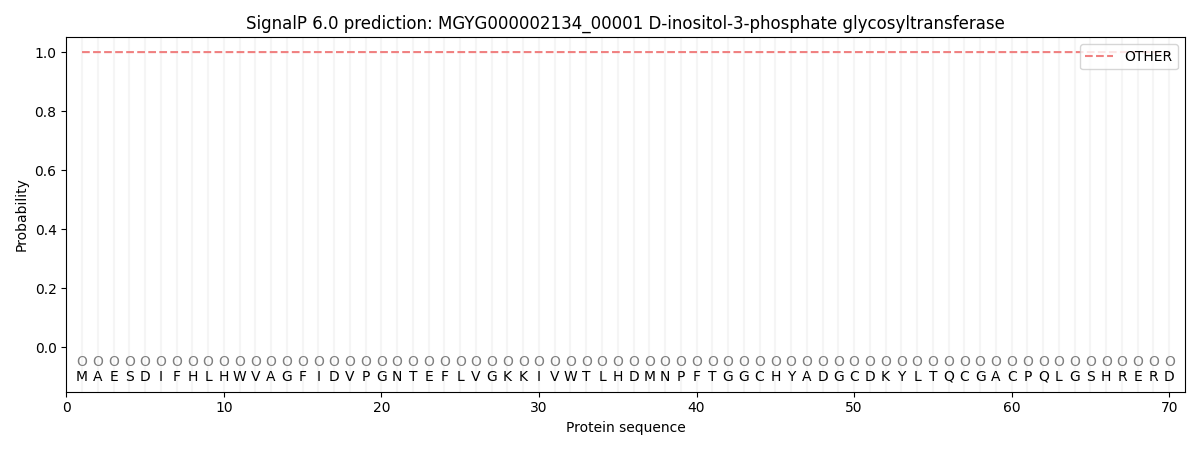
This protein is predicted as OTHER
| Other | SP_Sec_SPI | LIPO_Sec_SPII | TAT_Tat_SPI | TATLIP_Sec_SPII | PILIN_Sec_SPIII |
|---|---|---|---|---|---|
| 1.000050 | 0.000000 | 0.000000 | 0.000000 | 0.000000 | 0.000000 |