You are browsing environment: HUMAN GUT
CAZyme Information: MGYG000002328_01229
You are here: Home > Sequence: MGYG000002328_01229
Basic Information |
Genomic context |
Full Sequence |
Enzyme annotations |
CAZy signature domains |
CDD domains |
CAZyme hits |
PDB hits |
Swiss-Prot hits |
SignalP and Lipop annotations |
TMHMM annotations
Basic Information help
| Species | Enterocloster asparagiformis | |||||||||||
|---|---|---|---|---|---|---|---|---|---|---|---|---|
| Lineage | Bacteria; Firmicutes_A; Clostridia; Lachnospirales; Lachnospiraceae; Enterocloster; Enterocloster asparagiformis | |||||||||||
| CAZyme ID | MGYG000002328_01229 | |||||||||||
| CAZy Family | GT2 | |||||||||||
| CAZyme Description | hypothetical protein | |||||||||||
| CAZyme Property |
|
|||||||||||
| Genome Property |
|
|||||||||||
| Gene Location | Start: 18703; End: 20121 Strand: - | |||||||||||
CAZyme Signature Domains help
| Family | Start | End | Evalue | family coverage |
|---|---|---|---|---|
| GT2 | 5 | 171 | 5e-38 | 0.9823529411764705 |
CDD Domains download full data without filtering help
| Cdd ID | Domain | E-Value | qStart | qEnd | sStart | sEnd | Domain Description |
|---|---|---|---|---|---|---|---|
| cd10032 | UDG-F6_HDG | 1.16e-67 | 298 | 435 | 2 | 141 | Uracil DNA glycosylase family 6, includes hypoxanthine-DNA glycosylase and similar proteins. Uracil DNA glycosylase family 6 hypoxanthine-DNA glycosylase (HDG) lacks any detectable UDG activity; it excises hypoxanthine, a deamination product of adenine, from double-stranded DNA. Uracil-DNA glycosylase (UDGs) initiates repair of uracils in DNA. Uracil in DNA can arise as a result of misincorporation of dUMP residues by DNA polymerase or via deamination of cytosine. Uracil in DNA mispaired with guanine is one of the major pro-mutagenic events, causing G:C->A:T mutations. Thus, UDG is an essential enzyme for maintaining the integrity of genetic information. UDGs have been classified into various families on the basis of their substrate specificity, conserved motifs, and structural similarities. Although these families demonstrate different substrate specificities, often the function of one enzyme can be complemented by the other. |
| TIGR04274 | hypoxanDNAglyco | 2.80e-41 | 299 | 446 | 2 | 150 | hypoxanthine-DNA glycosylase. Members of this protein family represent family 6 of the uracil-DNA glycosylase superfamily, where the five previously described families all act as uracil-DNA glycosylase (EC 3.2.2.27) per se. This family, instead, acts as a hypoxanthine-DNA glycosylase, where hypoxanthine results from deamination of adenine. Activity was shown directly for members from Methanosarcina barkeri and Methanosarcina acetivorans. |
| pfam00535 | Glycos_transf_2 | 1.68e-39 | 5 | 169 | 1 | 164 | Glycosyl transferase family 2. Diverse family, transferring sugar from UDP-glucose, UDP-N-acetyl- galactosamine, GDP-mannose or CDP-abequose, to a range of substrates including cellulose, dolichol phosphate and teichoic acids. |
| cd00761 | Glyco_tranf_GTA_type | 1.76e-33 | 7 | 116 | 2 | 113 | Glycosyltransferase family A (GT-A) includes diverse families of glycosyl transferases with a common GT-A type structural fold. Glycosyltransferases (GTs) are enzymes that synthesize oligosaccharides, polysaccharides, and glycoconjugates by transferring the sugar moiety from an activated nucleotide-sugar donor to an acceptor molecule, which may be a growing oligosaccharide, a lipid, or a protein. Based on the stereochemistry of the donor and acceptor molecules, GTs are classified as either retaining or inverting enzymes. To date, all GT structures adopt one of two possible folds, termed GT-A fold and GT-B fold. This hierarchy includes diverse families of glycosyl transferases with a common GT-A type structural fold, which has two tightly associated beta/alpha/beta domains that tend to form a continuous central sheet of at least eight beta-strands. The majority of the proteins in this superfamily are Glycosyltransferase family 2 (GT-2) proteins. But it also includes families GT-43, GT-6, GT-8, GT13 and GT-7; which are evolutionarily related to GT-2 and share structure similarities. |
| COG3663 | Mug | 1.60e-30 | 298 | 446 | 1 | 157 | G:T/U-mismatch repair DNA glycosylase [Replication, recombination and repair]. |
CAZyme Hits help
| Hit ID | E-Value | Query Start | Query End | Hit Start | Hit End |
|---|---|---|---|---|---|
| QJU21468.1 | 1.70e-264 | 1 | 446 | 1 | 446 |
| ASN94231.1 | 5.67e-263 | 1 | 446 | 1 | 446 |
| QRP41091.1 | 5.67e-263 | 1 | 446 | 1 | 446 |
| QIX92878.1 | 3.12e-260 | 1 | 446 | 1 | 446 |
| ANU48636.1 | 3.63e-259 | 1 | 446 | 1 | 446 |
PDB Hits download full data without filtering help
| Hit ID | E-Value | Query Start | Query End | Hit Start | Hit End | Description |
|---|---|---|---|---|---|---|
| 5HEA_A | 6.54e-29 | 3 | 219 | 6 | 225 | CgTstructure in hexamer [Streptococcus parasanguinis FW213],5HEA_B CgT structure in hexamer [Streptococcus parasanguinis FW213],5HEA_C CgT structure in hexamer [Streptococcus parasanguinis FW213],5HEC_A CgT structure in dimer [Streptococcus parasanguinis FW213],5HEC_B CgT structure in dimer [Streptococcus parasanguinis FW213] |
| 3BCV_A | 5.18e-25 | 1 | 209 | 4 | 224 | Crystalstructure of a putative glycosyltransferase from Bacteroides fragilis [Bacteroides fragilis NCTC 9343],3BCV_B Crystal structure of a putative glycosyltransferase from Bacteroides fragilis [Bacteroides fragilis NCTC 9343] |
| 5TZE_C | 1.21e-21 | 5 | 221 | 4 | 215 | Crystalstructure of S. aureus TarS in complex with UDP-GlcNAc [Staphylococcus aureus],5TZE_E Crystal structure of S. aureus TarS in complex with UDP-GlcNAc [Staphylococcus aureus],5TZI_C Crystal structure of S. aureus TarS 1-349 [Staphylococcus aureus],5TZJ_A Crystal structure of S. aureus TarS 1-349 in complex with UDP-GlcNAc [Staphylococcus aureus],5TZJ_C Crystal structure of S. aureus TarS 1-349 in complex with UDP-GlcNAc [Staphylococcus aureus],5TZK_C Crystal structure of S. aureus TarS 1-349 in complex with UDP [Staphylococcus aureus] |
| 5TZ8_A | 4.52e-21 | 5 | 221 | 4 | 215 | Crystalstructure of S. aureus TarS [Staphylococcus aureus],5TZ8_B Crystal structure of S. aureus TarS [Staphylococcus aureus],5TZ8_C Crystal structure of S. aureus TarS [Staphylococcus aureus] |
| 2Z87_A | 6.80e-21 | 1 | 143 | 373 | 505 | Crystalstructure of chondroitin polymerase from Escherichia coli strain K4 (K4CP) complexed with UDP-GalNAc and UDP [Escherichia coli],2Z87_B Crystal structure of chondroitin polymerase from Escherichia coli strain K4 (K4CP) complexed with UDP-GalNAc and UDP [Escherichia coli] |
Swiss-Prot Hits download full data without filtering help
| Hit ID | E-Value | Query Start | Query End | Hit Start | Hit End | Description |
|---|---|---|---|---|---|---|
| P71059 | 7.82e-40 | 1 | 224 | 2 | 226 | Uncharacterized glycosyltransferase EpsJ OS=Bacillus subtilis (strain 168) OX=224308 GN=epsJ PE=2 SV=1 |
| P71057 | 9.17e-33 | 2 | 259 | 4 | 259 | Putative glycosyltransferase EpsH OS=Bacillus subtilis (strain 168) OX=224308 GN=epsH PE=2 SV=1 |
| A0A0H2URH7 | 9.84e-33 | 4 | 314 | 7 | 309 | Glycosyltransferase GlyA OS=Streptococcus pneumoniae serotype 4 (strain ATCC BAA-334 / TIGR4) OX=170187 GN=glyA PE=3 SV=1 |
| P11290 | 2.66e-23 | 4 | 198 | 8 | 210 | Uncharacterized glycosyltransferase YibD OS=Escherichia coli (strain K12) OX=83333 GN=yibD PE=3 SV=2 |
| A0A0H2UR96 | 1.17e-22 | 3 | 221 | 4 | 226 | Glycosyltransferase GlyG OS=Streptococcus pneumoniae serotype 4 (strain ATCC BAA-334 / TIGR4) OX=170187 GN=glyG PE=1 SV=1 |
SignalP and Lipop Annotations help
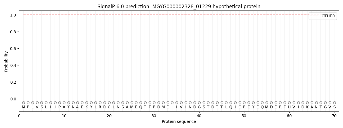
This protein is predicted as OTHER
| Other | SP_Sec_SPI | LIPO_Sec_SPII | TAT_Tat_SPI | TATLIP_Sec_SPII | PILIN_Sec_SPIII |
|---|---|---|---|---|---|
| 1.000048 | 0.000000 | 0.000000 | 0.000000 | 0.000000 | 0.000000 |