You are browsing environment: HUMAN GUT
CAZyme Information: MGYG000002341_02511
You are here: Home > Sequence: MGYG000002341_02511
Basic Information |
Genomic context |
Full Sequence |
Enzyme annotations |
CAZy signature domains |
CDD domains |
CAZyme hits |
PDB hits |
Swiss-Prot hits |
SignalP and Lipop annotations |
TMHMM annotations
Basic Information help
| Species | Acinetobacter ursingii | |||||||||||
|---|---|---|---|---|---|---|---|---|---|---|---|---|
| Lineage | Bacteria; Proteobacteria; Gammaproteobacteria; Pseudomonadales; Moraxellaceae; Acinetobacter; Acinetobacter ursingii | |||||||||||
| CAZyme ID | MGYG000002341_02511 | |||||||||||
| CAZy Family | CBM5 | |||||||||||
| CAZyme Description | Extracellular serine proteinase | |||||||||||
| CAZyme Property |
|
|||||||||||
| Genome Property |
|
|||||||||||
| Gene Location | Start: 24542; End: 25684 Strand: + | |||||||||||
CDD Domains download full data without filtering help
| Cdd ID | Domain | E-Value | qStart | qEnd | sStart | sEnd | Domain Description |
|---|---|---|---|---|---|---|---|
| cd04077 | Peptidases_S8_PCSK9_ProteinaseK_like | 5.02e-123 | 110 | 360 | 1 | 254 | Peptidase S8 family domain in ProteinaseK-like proteins. The peptidase S8 or Subtilase clan of proteases have a Asp/His/Ser catalytic triad that is not homologous to trypsin. This CD contains several members of this clan including: PCSK9 (Proprotein convertase subtilisin/kexin type 9), Proteinase_K, Proteinase_T, and other subtilisin-like serine proteases. PCSK9 posttranslationally regulates hepatic low-density lipoprotein receptors (LDLRs) by binding to LDLRs on the cell surface, leading to their degradation. The binding site of PCSK9 has been localized to the epidermal growth factor-like repeat A (EGF-A) domain of the LDLR. Characterized Proteinases K are secreted endopeptidases with a high degree of sequence conservation. Proteinases K are not substrate-specific and function in a wide variety of species in different pathways. It can hydrolyze keratin and other proteins with subtilisin-like specificity. The number of calcium-binding motifs found in these differ. Proteinase T is a novel proteinase from the fungus Tritirachium album Limber. The amino acid sequence of proteinase T as deduced from the nucleotide sequence is about 56% identical to that of proteinase K. |
| cd07484 | Peptidases_S8_Thermitase_like | 1.13e-61 | 134 | 341 | 29 | 239 | Peptidase S8 family domain in Thermitase-like proteins. Thermitase is a non-specific, trypsin-related serine protease with a very high specific activity. It contains a subtilisin like domain. The tertiary structure of thermitase is similar to that of subtilisin BPN'. It contains a Asp/His/Ser catalytic triad. Members of the peptidases S8 (subtilisin and kexin) and S53 (sedolisin) clan include endopeptidases and exopeptidases. The S8 family has an Asp/His/Ser catalytic triad similar to that found in trypsin-like proteases, but do not share their three-dimensional structure and are not homologous to trypsin. Serine acts as a nucleophile, aspartate as an electrophile, and histidine as a base. The S53 family contains a catalytic triad Glu/Asp/Ser with an additional acidic residue Asp in the oxyanion hole, similar to that of subtilisin. The serine residue here is the nucleophilic equivalent of the serine residue in the S8 family, while glutamic acid has the same role here as the histidine base. However, the aspartic acid residue that acts as an electrophile is quite different. In S53 the it follows glutamic acid, while in S8 it precedes histidine. The stability of these enzymes may be enhanced by calcium, some members have been shown to bind up to 4 ions via binding sites with different affinity. There is a great diversity in the characteristics of their members: some contain disulfide bonds, some are intracellular while others are extracellular, some function at extreme temperatures, and others at high or low pH values. |
| cd07477 | Peptidases_S8_Subtilisin_subset | 3.29e-55 | 134 | 355 | 1 | 226 | Peptidase S8 family domain in Subtilisin proteins. This group is composed of many different subtilisins: Pro-TK-subtilisin, subtilisin Carlsberg, serine protease Pb92 subtilisin, and BPN subtilisins just to name a few. Pro-TK-subtilisin is a serine protease from the hyperthermophilic archaeon Thermococcus kodakaraensis and consists of a signal peptide, a propeptide, and a mature domain. TK-subtilisin is matured from pro-TK-subtilisin upon autoprocessing and degradation of the propeptide. Unlike other subtilisins though, the folding of the unprocessed form of pro-TK-subtilisin is induced by Ca2+ binding which is almost completed prior to autoprocessing. Ca2+ is required for activity unlike the bacterial subtilisins. The propeptide is not required for folding of the mature domain unlike the bacterial subtilases because of the stability produced from Ca2+ binding. Subtilisin Carlsberg is extremely similar in structure to subtilisin BPN'/Novo thought it has a 30% difference in amino acid sequence. The substrate binding regions are also similar and 2 possible Ca2+ binding sites have been identified recently. Subtilisin Carlsberg possesses the highest commercial importance as a proteolytic additive for detergents. Serine protease Pb92, the serine protease from the alkalophilic Bacillus strain PB92, also contains two calcium ions and the overall folding of the polypeptide chain closely resembles that of the subtilisins. Members of the peptidases S8 and S35 clan include endopeptidases, exopeptidases and also a tripeptidyl-peptidase. The S8 family has an Asp/His/Ser catalytic triad similar to that found in trypsin-like proteases, but do not share their three-dimensional structure and are not homologous to trypsin. The S53 family contains a catalytic triad Glu/Asp/Ser. The stability of these enzymes may be enhanced by calcium, some members have been shown to bind up to 4 ions via binding sites with different affinity. Some members of this clan contain disulfide bonds. These enzymes can be intra- and extracellular, some function at extreme temperatures and pH values. |
| cd00306 | Peptidases_S8_S53 | 3.75e-50 | 135 | 357 | 1 | 240 | Peptidase domain in the S8 and S53 families. Members of the peptidases S8 (subtilisin and kexin) and S53 (sedolisin) family include endopeptidases and exopeptidases. The S8 family has an Asp/His/Ser catalytic triad similar to that found in trypsin-like proteases, but do not share their three-dimensional structure and are not homologous to trypsin. Serine acts as a nucleophile, aspartate as an electrophile, and histidine as a base. The S53 family contains a catalytic triad Glu/Asp/Ser with an additional acidic residue Asp in the oxyanion hole, similar to that of subtilisin. The serine residue here is the nucleophilic equivalent of the serine residue in the S8 family, while glutamic acid has the same role here as the histidine base. However, the aspartic acid residue that acts as an electrophile is quite different. In S53, it follows glutamic acid, while in S8 it precedes histidine. The stability of these enzymes may be enhanced by calcium; some members have been shown to bind up to 4 ions via binding sites with different affinity. There is a great diversity in the characteristics of their members: some contain disulfide bonds, some are intracellular while others are extracellular, some function at extreme temperatures, and others at high or low pH values. |
| cd07487 | Peptidases_S8_1 | 3.11e-49 | 133 | 360 | 2 | 264 | Peptidase S8 family domain, uncharacterized subfamily 1. This family is a member of the Peptidases S8 or Subtilases serine endo- and exo-peptidase clan. They have an Asp/His/Ser catalytic triad similar to that found in trypsin-like proteases, but do not share their three-dimensional structure and are not homologous to trypsin. The stability of subtilases may be enhanced by calcium, some members have been shown to bind up to 4 ions via binding sites with different affinity. Some members of this clan contain disulfide bonds. These enzymes can be intra- and extracellular, some function at extreme temperatures and pH values. |
CAZyme Hits help
| Hit ID | E-Value | Query Start | Query End | Hit Start | Hit End |
|---|---|---|---|---|---|
| QOC24262.1 | 1.09e-110 | 52 | 377 | 57 | 378 |
| ADO75815.1 | 2.57e-91 | 60 | 377 | 86 | 398 |
| QRK11067.1 | 7.71e-89 | 54 | 376 | 81 | 403 |
| AXO37891.1 | 3.98e-78 | 54 | 377 | 52 | 374 |
| AEB45483.1 | 4.69e-77 | 54 | 377 | 81 | 403 |
PDB Hits download full data without filtering help
| Hit ID | E-Value | Query Start | Query End | Hit Start | Hit End | Description |
|---|---|---|---|---|---|---|
| 2B6N_A | 3.10e-107 | 105 | 377 | 3 | 275 | The1.8 A crystal structure of a Proteinase K like enzyme from a psychrotroph Serratia species [Serratia sp. GF96] |
| 4DZT_A | 3.81e-105 | 104 | 380 | 2 | 276 | AqualysinI: the crystal structure of a serine protease from an extreme thermophile, Thermus aquaticus YT-1 [Thermus aquaticus] |
| 5WSL_A | 1.53e-102 | 104 | 377 | 2 | 275 | Structuralstudies of keratinase from Meiothermus taiwanensis WR-220 [Meiothermus taiwanensis WR-220],5WSL_B Structural studies of keratinase from Meiothermus taiwanensis WR-220 [Meiothermus taiwanensis WR-220],5WSL_C Structural studies of keratinase from Meiothermus taiwanensis WR-220 [Meiothermus taiwanensis WR-220] |
| 1S2N_A | 7.28e-97 | 105 | 377 | 1 | 270 | Crystalstructure of a cold adapted subtilisin-like serine proteinase [Vibrio sp. PA-44],1S2N_B Crystal structure of a cold adapted subtilisin-like serine proteinase [Vibrio sp. PA-44],1SH7_A Crystal structure of a cold adapted subtilisin-like serine proteinase [Vibrio sp. PA-44],1SH7_B Crystal structure of a cold adapted subtilisin-like serine proteinase [Vibrio sp. PA-44] |
| 1BJR_E | 9.75e-68 | 105 | 342 | 3 | 240 | ComplexFormed Between Proteolytically Generated Lactoferrin Fragment And Proteinase K [Parengyodontium album],1CNM_A Enhancement Of Catalytic Efficiency Of Proteinase K Through Exposure To Anhydrous Organic Solvent At 70 Degrees Celsius [Parengyodontium album],1EGQ_A Enhancement Of Enzyme Activity Through Three-Phase Partitioning: Crystal Structure Of A Modified Serine Proteinase At 1.5 A Resolution [Parengyodontium album],1OYO_A Regulation of protease activity by melanin: Crystal structure of the complex formed between proteinase K and melanin monomers at 2.0 resolution [Parengyodontium album],1PJ8_A Structure of a ternary complex of proteinase K, mercury and a substrate-analogue hexapeptide at 2.2 A resolution [Parengyodontium album],1PTK_A Studies On The Inhibitory Action Of Mercury Upon Proteinase K [Parengyodontium album],2DP4_E Crystal structure of the complex formed between proteinase K and a human lactoferrin fragment at 2.9 A resolution [Parengyodontium album],2PKC_A CRYSTAL STRUCTURE OF CALCIUM-FREE PROTEINASE K AT 1.5 ANGSTROMS RESOLUTION [Parengyodontium album],2PRK_A SYNCHROTRON X-RAY DATA COLLECTION AND RESTRAINED LEAST-SQUARES REFINEMENT OF THE CRYSTAL STRUCTURE OF PROTEINASE K AT 1.5 ANGSTROMS RESOLUTION [Parengyodontium album],3D9Q_X Proteinase K by LB nanotemplate method before high X-Ray dose on ESRF ID23-1 beamline [Parengyodontium album],3DDZ_X Proteinase K by LB nanotemplate method after the first step of high X-Ray dose on ESRF ID23-1 beamline [Parengyodontium album],3DE0_X Proteinase K by LB nanotemplate method after the second step of high X-Ray dose on ESRF ID23-1 beamline [Parengyodontium album],3DE1_X Proteinase K by LB nanotemplate method after the third step of high X-Ray dose on ESRF ID23-1 beamline [Parengyodontium album],3DE2_X Proteinase K by LB nanotemplate method after the fourth step of high X-Ray dose on ESRF ID23-1 beamline [Parengyodontium album],3DE3_X Proteinase K by Classical hanging drop method before high X-Ray dose on ESRF ID23-1 beamline [Parengyodontium album],3DE4_X Proteinase K by Classical hanging drop method after the first step of high X-Ray dose on ESRF ID23-1 beamline [Parengyodontium album],3DE5_X roteinase K by Classical hanging drop method after the second step of high X-Ray dose on ESRF ID23-1 beamline [Parengyodontium album],3DE6_X Proteinase K by Classical hanging drop method after the third step of high X-Ray dose on ESRF ID23-1 beamline [Parengyodontium album],3DE7_X Proteinase K by Classical hanging drop method after the fourth step of high X-Ray dose on ESRF ID23-1 beamline [Parengyodontium album],3DVQ_X Proteinase K by LB nanotemplate method before high X-Ray dose on ESRF ID14-2 beamline [Parengyodontium album],3DVR_X Proteinase K by LB nanotemplate method after the first step of high X-Ray dose on ESRF ID14-2 beamline [Parengyodontium album],3DVS_X Proteinase K by LB nanotmplate method after the second step of high dose on ESRF ID14-2 beamline [Parengyodontium album],3DW1_X Proteinase K by LB nanotemplate method after the third step high X-Ray dose on ESRF ID14-2 beamline [Parengyodontium album],3DW3_X Proteinase K by Classical hanging drop method before high X Ray dose on ESRF ID 14-2 beamline [Parengyodontium album],3DWE_X Proteinase K by Classical hanging drop method after high X-Ray dose on ESRF ID14-2 beamline [Parengyodontium album],3I2Y_X Proteinase K by Classical hanging drop Method before high X-Ray dose on ID14-2 Beamline at ESRF [Parengyodontium album],3I30_X Proteinase K by Classical hanging drop Method after high X-Ray dose on ID14-2 Beamline at ESRF [Parengyodontium album],3I34_X Proteinase K by LB Nanotemplate Method after high X-Ray dose on ID14-2 Beamline at ESRF [Parengyodontium album],3I37_X Proteinase K by LB Nanotemplate Method before high X-Ray dose on ID14-2 Beamline at ESRF [Parengyodontium album],3PRK_E INHIBITION OF PROTEINASE K BY METHOXYSUCCINYL-ALA-ALA-PRO-ALA-CHLOROMETHYL KETONE. AN X-RAY STUDY AT 2.2-ANGSTROMS RESOLUTION [Parengyodontium album],3PTL_A Crystal structure of proteinase K inhibited by a lactoferrin nonapeptide, Lys-Gly-Glu-Ala-Asp-Ala-Leu-Ser-Leu-Asp at 1.3 A resolution. [Parengyodontium album],4DJ5_X Proteinase K by Langmuir-Blodgett Hanging Drop Method at 1.8A resolution for Unique Water Distribution [Parengyodontium album],5AMX_A Crystal Structure of Proteinase K processed with the CrystalDirect automated mounting and cryo-cooling technology [Parengyodontium album],5K7S_A MicroED structure of proteinase K at 1.6 A resolution [Parengyodontium album],6CL7_A 1.71 A MicroED structure of proteinase K at 0.86 e- / A^2 [Parengyodontium album],6CL8_A 2.00 A MicroED structure of proteinase K at 2.6 e- / A^2 [Parengyodontium album],6CL9_A 2.20 A MicroED structure of proteinase K at 4.3 e- / A^2 [Parengyodontium album],6CLA_A 2.80 A MicroED structure of proteinase K at 6.0 e- / A^2 [Parengyodontium album],6CLB_A 3.20 A MicroED structure of proteinase K at 7.8 e- / A^2 [Parengyodontium album],6KKF_A Crystal structure of proteinase K complexed with a triglycine [Parengyodontium album],6PKJ_A MicroED structure of proteinase K from an uncoated, single lamella at 2.17A resolution (#2) [Parengyodontium album],6PKK_A MicroED structure of proteinase K from an uncoated, single lamella at 2.18A resolution (#5) [Parengyodontium album],6PKL_A MicroED structure of proteinase K from an uncoated, single lamella at 2.59A resolution (#7) [Parengyodontium album],6PKM_A MicroED structure of proteinase K from an uncoated, single lamella at 2.17A resolution (#8) [Parengyodontium album],6PKN_A MicroED structure of proteinase K from an unpolished, platinum-coated, single lamella at 2.08A resolution (#9) [Parengyodontium album],6PKO_A MicroED structure of proteinase K from a platinum coated, unpolished, single lamella at 2.07A resolution (#12) [Parengyodontium album],6PKP_A MicroED structure of proteinase K from a platinum-coated, polished, single lamella at 1.91A resolution (#10) [Parengyodontium album],6PKQ_A MicroED structure of proteinase K from a platinum-coated, polished, single lamella at 1.85A resolution (#11) [Parengyodontium album],6PKR_A MicroED structure of proteinase K from a platinum-coated, polished, single lamella at 1.79A resolution (#13) [Parengyodontium album],6PKS_A MicroED structure of proteinase K from low-dose merged lamellae that were not pre-coated with platinum 2.16A resolution (LD) [Parengyodontium album],6PKT_A MicroED structure of proteinase K from merging low-dose, platinum pre-coated lamellae at 1.85A resolution (LDPT) [Parengyodontium album],6PQ0_A LCP-embedded Proteinase K treated with MPD [Parengyodontium album],6PQ4_A LCP-embedded Proteinase K treated with lipase [Parengyodontium album],6PU4_A MicroED structure of proteinase K recorded on Falcon III [Parengyodontium album],6PU5_A MicroED structure of proteinase K recorded on CetaD [Parengyodontium album],7JSY_A Chain A, Proteinase K [Parengyodontium album] |
Swiss-Prot Hits download full data without filtering help
| Hit ID | E-Value | Query Start | Query End | Hit Start | Hit End | Description |
|---|---|---|---|---|---|---|
| P80146 | 4.68e-118 | 55 | 378 | 90 | 408 | Extracellular serine proteinase OS=Thermus sp. (strain Rt41A) OX=32063 PE=1 SV=3 |
| P08594 | 9.87e-107 | 60 | 380 | 90 | 403 | Aqualysin-1 OS=Thermus aquaticus OX=271 GN=pstI PE=1 SV=2 |
| P16588 | 1.40e-96 | 55 | 377 | 93 | 413 | Alkaline serine exoprotease A OS=Vibrio alginolyticus OX=663 GN=proA PE=3 SV=1 |
| P06873 | 1.28e-77 | 30 | 342 | 40 | 345 | Proteinase K OS=Parengyodontium album OX=37998 GN=PROK PE=1 SV=2 |
| R4IR27 | 2.87e-77 | 54 | 377 | 62 | 382 | Subtilisin-like serine protease AsES OS=Sarocladium strictum OX=5046 GN=AsES PE=1 SV=1 |
SignalP and Lipop Annotations help
This protein is predicted as SP
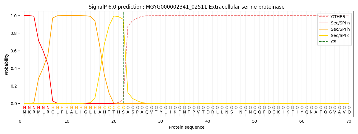
| Other | SP_Sec_SPI | LIPO_Sec_SPII | TAT_Tat_SPI | TATLIP_Sec_SPII | PILIN_Sec_SPIII |
|---|---|---|---|---|---|
| 0.000442 | 0.998792 | 0.000226 | 0.000197 | 0.000176 | 0.000174 |