You are browsing environment: HUMAN GUT
CAZyme Information: MGYG000002383_02751
You are here: Home > Sequence: MGYG000002383_02751
Basic Information |
Genomic context |
Full Sequence |
Enzyme annotations |
CAZy signature domains |
CDD domains |
CAZyme hits |
PDB hits |
Swiss-Prot hits |
SignalP and Lipop annotations |
TMHMM annotations
Basic Information help
| Species | Lacticaseibacillus zeae | |||||||||||
|---|---|---|---|---|---|---|---|---|---|---|---|---|
| Lineage | Bacteria; Firmicutes; Bacilli; Lactobacillales; Lactobacillaceae; Lacticaseibacillus; Lacticaseibacillus zeae | |||||||||||
| CAZyme ID | MGYG000002383_02751 | |||||||||||
| CAZy Family | GH13 | |||||||||||
| CAZyme Description | Oligo-1,6-glucosidase | |||||||||||
| CAZyme Property |
|
|||||||||||
| Genome Property |
|
|||||||||||
| Gene Location | Start: 141107; End: 142678 Strand: + | |||||||||||
CAZyme Signature Domains help
| Family | Start | End | Evalue | family coverage |
|---|---|---|---|---|
| GH13 | 1 | 344 | 1e-161 | 0.9684813753581661 |
CDD Domains download full data without filtering help
| Cdd ID | Domain | E-Value | qStart | qEnd | sStart | sEnd | Domain Description |
|---|---|---|---|---|---|---|---|
| TIGR02403 | trehalose_treC | 0.0 | 1 | 523 | 33 | 543 | alpha,alpha-phosphotrehalase. Trehalose is a glucose disaccharide that serves in many biological systems as a compatible solute for protection against hyperosmotic and thermal stress. This family describes trehalose-6-phosphate hydrolase, product of the treC (or treA) gene, which is often found together with a trehalose uptake transporter and a trehalose operon repressor. |
| PRK10933 | PRK10933 | 0.0 | 1 | 518 | 39 | 545 | trehalose-6-phosphate hydrolase; Provisional |
| cd11333 | AmyAc_SI_OligoGlu_DGase | 0.0 | 2 | 443 | 32 | 427 | Alpha amylase catalytic domain found in Sucrose isomerases, oligo-1,6-glucosidase (also called isomaltase; sucrase-isomaltase; alpha-limit dextrinase), dextran glucosidase (also called glucan 1,6-alpha-glucosidase), and related proteins. The sucrose isomerases (SIs) Isomaltulose synthase (EC 5.4.99.11) and Trehalose synthase (EC 5.4.99.16) catalyze the isomerization of sucrose and maltose to produce isomaltulose and trehalulose, respectively. Oligo-1,6-glucosidase (EC 3.2.1.10) hydrolyzes the alpha-1,6-glucosidic linkage of isomaltooligosaccharides, pannose, and dextran. Unlike alpha-1,4-glucosidases (EC 3.2.1.20), it fails to hydrolyze the alpha-1,4-glucosidic bonds of maltosaccharides. Dextran glucosidase (DGase, EC 3.2.1.70) hydrolyzes alpha-1,6-glucosidic linkages at the non-reducing end of panose, isomaltooligosaccharides and dextran to produce alpha-glucose.The common reaction chemistry of the alpha-amylase family enzymes is based on a two-step acid catalytic mechanism that requires two critical carboxylates: one acting as a general acid/base (Glu) and the other as a nucleophile (Asp). Both hydrolysis and transglycosylation proceed via the nucleophilic substitution reaction between the anomeric carbon, C1 and a nucleophile. Both enzymes contain the three catalytic residues (Asp, Glu and Asp) common to the alpha-amylase family as well as two histidine residues which are predicted to be critical to binding the glucose residue adjacent to the scissile bond in the substrates. The Alpha-amylase family comprises the largest family of glycoside hydrolases (GH), with the majority of enzymes acting on starch, glycogen, and related oligo- and polysaccharides. These proteins catalyze the transformation of alpha-1,4 and alpha-1,6 glucosidic linkages with retention of the anomeric center. The protein is described as having 3 domains: A, B, C. A is a (beta/alpha) 8-barrel; B is a loop between the beta 3 strand and alpha 3 helix of A; C is the C-terminal extension characterized by a Greek key. The majority of the enzymes have an active site cleft found between domains A and B where a triad of catalytic residues performs catalysis. Other members of this family have lost the catalytic activity as in the case of the human 4F2hc, or only have 2 residues that serve as the catalytic nucleophile and the acid/base, such as Thermus A4 beta-galactosidase with 2 Glu residues (GH42) and human alpha-galactosidase with 2 Asp residues (GH31). The family members are quite extensive and include: alpha amylase, maltosyltransferase, cyclodextrin glycotransferase, maltogenic amylase, neopullulanase, isoamylase, 1,4-alpha-D-glucan maltotetrahydrolase, 4-alpha-glucotransferase, oligo-1,6-glucosidase, amylosucrase, sucrose phosphorylase, and amylomaltase. |
| pfam00128 | Alpha-amylase | 8.57e-143 | 2 | 348 | 11 | 334 | Alpha amylase, catalytic domain. Alpha amylase is classified as family 13 of the glycosyl hydrolases. The structure is an 8 stranded alpha/beta barrel containing the active site, interrupted by a ~70 a.a. calcium-binding domain protruding between beta strand 3 and alpha helix 3, and a carboxyl-terminal Greek key beta-barrel domain. |
| COG0366 | AmyA | 6.32e-137 | 2 | 499 | 36 | 499 | Glycosidase [Carbohydrate transport and metabolism]. |
CAZyme Hits help
| Hit ID | E-Value | Query Start | Query End | Hit Start | Hit End |
|---|---|---|---|---|---|
| QEW11664.1 | 0.0 | 1 | 523 | 36 | 558 |
| QQN31974.1 | 0.0 | 1 | 523 | 36 | 558 |
| QFV11669.1 | 0.0 | 1 | 523 | 36 | 558 |
| AGP70261.1 | 0.0 | 1 | 523 | 36 | 558 |
| QJZ29180.1 | 0.0 | 1 | 523 | 36 | 558 |
PDB Hits download full data without filtering help
| Hit ID | E-Value | Query Start | Query End | Hit Start | Hit End | Description |
|---|---|---|---|---|---|---|
| 1UOK_A | 4.03e-201 | 1 | 522 | 37 | 556 | CrystalStructure Of B. Cereus Oligo-1,6-Glucosidase [Bacillus cereus] |
| 5DO8_A | 9.67e-199 | 1 | 522 | 38 | 551 | 1.8Angstrom crystal structure of Listeria monocytogenes Lmo0184 alpha-1,6-glucosidase [Listeria monocytogenes EGD-e],5DO8_B 1.8 Angstrom crystal structure of Listeria monocytogenes Lmo0184 alpha-1,6-glucosidase [Listeria monocytogenes EGD-e],5DO8_C 1.8 Angstrom crystal structure of Listeria monocytogenes Lmo0184 alpha-1,6-glucosidase [Listeria monocytogenes EGD-e] |
| 4M56_A | 3.01e-176 | 1 | 518 | 36 | 555 | TheStructure of Wild-type MalL from Bacillus subtilis [Bacillus subtilis subsp. subtilis str. 168],4M56_B The Structure of Wild-type MalL from Bacillus subtilis [Bacillus subtilis subsp. subtilis str. 168] |
| 4MB1_A | 6.04e-176 | 1 | 518 | 36 | 555 | TheStructure of MalL mutant enzyme G202P from Bacillus subtilus [Bacillus subtilis subsp. subtilis str. 168] |
| 5WCZ_A | 6.90e-176 | 1 | 518 | 61 | 580 | CrystalStructure of Wild-Type MalL from Bacillus subtilis with TS analogue 1-deoxynojirimycin [Bacillus subtilis subsp. subtilis str. 168],5WCZ_B Crystal Structure of Wild-Type MalL from Bacillus subtilis with TS analogue 1-deoxynojirimycin [Bacillus subtilis subsp. subtilis str. 168] |
Swiss-Prot Hits download full data without filtering help
| Hit ID | E-Value | Query Start | Query End | Hit Start | Hit End | Description |
|---|---|---|---|---|---|---|
| P29094 | 2.32e-213 | 1 | 522 | 37 | 558 | Oligo-1,6-glucosidase OS=Parageobacillus thermoglucosidasius OX=1426 GN=malL PE=1 SV=1 |
| Q9K8U9 | 5.24e-207 | 1 | 521 | 37 | 556 | Oligo-1,6-glucosidase OS=Alkalihalobacillus halodurans (strain ATCC BAA-125 / DSM 18197 / FERM 7344 / JCM 9153 / C-125) OX=272558 GN=malL PE=3 SV=1 |
| P43473 | 1.85e-201 | 1 | 522 | 39 | 555 | Alpha-glucosidase OS=Pediococcus pentosaceus OX=1255 GN=agl PE=3 SV=1 |
| P21332 | 2.21e-200 | 1 | 522 | 37 | 556 | Oligo-1,6-glucosidase OS=Bacillus cereus OX=1396 GN=malL PE=1 SV=1 |
| Q45101 | 8.46e-182 | 1 | 521 | 36 | 551 | Oligo-1,6-glucosidase OS=Weizmannia coagulans OX=1398 GN=malL PE=3 SV=1 |
SignalP and Lipop Annotations help
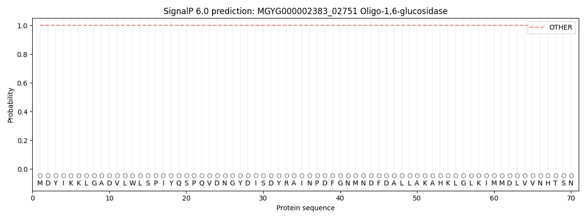
This protein is predicted as OTHER
| Other | SP_Sec_SPI | LIPO_Sec_SPII | TAT_Tat_SPI | TATLIP_Sec_SPII | PILIN_Sec_SPIII |
|---|---|---|---|---|---|
| 1.000059 | 0.000000 | 0.000000 | 0.000000 | 0.000000 | 0.000000 |