You are browsing environment: HUMAN GUT
CAZyme Information: MGYG000002416_00770
You are here: Home > Sequence: MGYG000002416_00770
Basic Information |
Genomic context |
Full Sequence |
Enzyme annotations |
CAZy signature domains |
CDD domains |
CAZyme hits |
PDB hits |
Swiss-Prot hits |
SignalP and Lipop annotations |
TMHMM annotations
Basic Information help
| Species | Hydrogeniiclostridium mannosilyticum | |||||||||||
|---|---|---|---|---|---|---|---|---|---|---|---|---|
| Lineage | Bacteria; Firmicutes_A; Clostridia; Oscillospirales; Acutalibacteraceae; Hydrogeniiclostridium; Hydrogeniiclostridium mannosilyticum | |||||||||||
| CAZyme ID | MGYG000002416_00770 | |||||||||||
| CAZy Family | GH130 | |||||||||||
| CAZyme Description | Beta-1,4-mannooligosaccharide phosphorylase | |||||||||||
| CAZyme Property |
|
|||||||||||
| Genome Property |
|
|||||||||||
| Gene Location | Start: 831482; End: 832474 Strand: - | |||||||||||
CAZyme Signature Domains help
| Family | Start | End | Evalue | family coverage |
|---|---|---|---|---|
| GH130 | 25 | 323 | 3.1e-89 | 0.9932432432432432 |
CDD Domains download full data without filtering help
| Cdd ID | Domain | E-Value | qStart | qEnd | sStart | sEnd | Domain Description |
|---|---|---|---|---|---|---|---|
| cd08993 | GH130 | 3.62e-158 | 40 | 320 | 1 | 279 | Glycosyl hydrolase family 130. This subfamily contains glycosyl hydrolase family 130 (GH130) proteins, as classified by the carbohydrate-active enzymes database (CAZY), are phosphorylases and hydrolases for beta-mannosides, and include beta-1,4-mannosylglucose phosphorylase (EC 2.4.1.281), beta-1,4-mannooligosaccharide phosphorylase (EC 2.4.1.319), among others that have yet to be characterized. They possess 5-bladed beta-propeller domains similar to families 32, 43, 62, 68, 117 (GH32, GH43, GH62, GH68, GH117). GH130 enzymes are involved in the bacterial utilization of mannans or N-linked glycans. Beta-1,4-mannosylglucose phosphorylase is involved in degradation of beta-1,4-D-mannosyl-N-acetyl-D-glucosamine linkages in the core of N-glycans; it produces alpha-mannose 1-phosphate and glucose from 4-O-beta-D-mannosyl-D-glucose and inorganic phosphate, using a critical catalytic Asp as a proton donor. This family includes Ruminococcus albus 4-O-beta-D-mannosyl-D-glucose phosphorylase (RaMP1) and beta-(1,4)-mannooligosaccharide phosphorylase (RaMP2), enzymes that phosphorolyze beta-mannosidic linkages at the non-reducing ends of their substrates, and have substantially diverse substrate specificity that are determined by three loop regions. |
| COG2152 | COG2152 | 6.58e-94 | 23 | 326 | 3 | 312 | Predicted glycosyl hydrolase, GH43/DUF377 family [Carbohydrate transport and metabolism]. |
| cd18615 | GH130 | 1.78e-86 | 43 | 314 | 6 | 277 | Glycosyl hydrolase family 130; uncharacterized. This subfamily contains glycosyl hydrolase family 130 (GH130) proteins, as classified by the carbohydrate-active enzymes database (CAZY), most of which are as yet uncharacterized. GH130 enzymes are phosphorylases and hydrolases for beta-mannosides, and include beta-1,4-mannosylglucose phosphorylase (EC 2.4.1.281), beta-1,4-mannooligosaccharide phosphorylase (EC 2.4.1.319), beta-1,4-mannosyl-N-acetyl-glucosamine phosphorylase (EC 2.4.1.320), beta-1,2-mannobiose phosphorylase (EC 2.4.1.-), beta-1,2-oligomannan phosphorylase (EC 2.4.1.-) and beta-1,2-mannosidase (EC 3.2.1.-). They possess 5-bladed beta-propeller domains similar to families 32, 43, 62, 68, 117 (GH32, GH43, GH62, GH68, GH117). GH130 enzymes are involved in the bacterial utilization of mannans or N-linked glycans. Beta-1,4-mannosylglucose phosphorylase is involved in degradation of beta-1,4-D-mannosyl-N-acetyl-D-glucosamine linkages in the core of N-glycans; it produces alpha-mannose 1-phosphate and glucose from 4-O-beta-D-mannosyl-D-glucose and inorganic phosphate, using a critical catalytic Asp as a proton donor. |
| cd18607 | GH130 | 4.79e-76 | 42 | 313 | 3 | 268 | Glycoside hydrolase family 130. Members of the glycosyl hydrolase family 130, as classified by the carbohydrate-active enzymes database (CAZY), are phosphorylases and hydrolases for beta-mannosides, and include beta-1,4-mannosylglucose phosphorylase (EC 2.4.1.281), beta-1,4-mannooligosaccharide phosphorylase (EC 2.4.1.319), beta-1,4-mannosyl-N-acetyl-glucosamine phosphorylase (EC 2.4.1.320), beta-1,2-mannobiose phosphorylase (EC 2.4.1.-), beta-1,2-oligomannan phosphorylase (EC 2.4.1.-) and beta-1,2-mannosidase (EC 3.2.1.-). They possess 5-bladed beta-propeller domains similar to families 32, 43, 62, 68, 117 (GH32, GH43, GH62, GH68, GH117). GH130 enzymes are involved in the bacterial utilization of mannans or N-linked glycans. Beta-1,4-mannosylglucose phosphorylase is involved in degradation of beta-1,4-D-mannosyl-N-acetyl-D-glucosamine linkages in the core of N-glycans; it produces alpha-mannose 1-phosphate and glucose from 4-O-beta-D-mannosyl-D-glucose and inorganic phosphate, using a critical catalytic Asp as a proton donor. |
| cd18612 | GH130_Lin0857-like | 1.06e-73 | 45 | 315 | 6 | 261 | Glycoside hydrolase family 130 such as Listeria innocua beta-1,2-mannobiose phosphorylase. This subfamily contains the glycosyl hydrolase family 130 (GH130), as classified by the carbohydrate-active enzymes database (CAZY), enzymes that are phosphorylases and hydrolases for beta-mannosides, and includes Listeria innocua beta-1,2-mannobiose phosphorylase (Lin0857). hey possess 5-bladed beta-propeller domains similar to families 32, 43, 62, 68, 117 (GH32, GH43, GH62, GH68, GH117). GH130 enzymes are involved in the bacterial utilization of mannans or N-linked glycans. Structure of Lin0857 shows beta-1,2-mannotriose bound in a U-shape, interacting with a phosphate analog at both ends. Lin0857 has a unique dimer structure connected by a loop, with a significant open-close loop displacement observed for substrate entry. A long loop, which is exclusively present in Lin0857, covers the active site to limit the pocket size. |
CAZyme Hits help
| Hit ID | E-Value | Query Start | Query End | Hit Start | Hit End |
|---|---|---|---|---|---|
| QUL53205.1 | 3.15e-201 | 1 | 325 | 1 | 325 |
| AIQ42764.1 | 9.02e-201 | 1 | 325 | 1 | 325 |
| AIQ31240.1 | 2.58e-200 | 1 | 325 | 1 | 325 |
| AIQ60001.1 | 3.67e-200 | 1 | 325 | 1 | 325 |
| AZK45217.1 | 4.50e-200 | 1 | 325 | 1 | 325 |
PDB Hits download full data without filtering help
| Hit ID | E-Value | Query Start | Query End | Hit Start | Hit End | Description |
|---|---|---|---|---|---|---|
| 4UDG_A | 2.75e-165 | 12 | 329 | 29 | 347 | Crystalstructure of b-1,4-mannopyranosyl-chitobiose phosphorylase at 1.60 Angstrom in complex with N-acetylglucosamine and inorganic phosphate [uncultured organism],4UDG_B Crystal structure of b-1,4-mannopyranosyl-chitobiose phosphorylase at 1.60 Angstrom in complex with N-acetylglucosamine and inorganic phosphate [uncultured organism],4UDG_C Crystal structure of b-1,4-mannopyranosyl-chitobiose phosphorylase at 1.60 Angstrom in complex with N-acetylglucosamine and inorganic phosphate [uncultured organism],4UDG_D Crystal structure of b-1,4-mannopyranosyl-chitobiose phosphorylase at 1.60 Angstrom in complex with N-acetylglucosamine and inorganic phosphate [uncultured organism],4UDG_E Crystal structure of b-1,4-mannopyranosyl-chitobiose phosphorylase at 1.60 Angstrom in complex with N-acetylglucosamine and inorganic phosphate [uncultured organism],4UDG_F Crystal structure of b-1,4-mannopyranosyl-chitobiose phosphorylase at 1.60 Angstrom in complex with N-acetylglucosamine and inorganic phosphate [uncultured organism],4UDI_A Crystal structure of b-1,4-mannopyranosyl-chitobiose phosphorylase at 1.85 Angstrom from unknown human gut bacteria (Uhgb_MP) [uncultured organism],4UDI_B Crystal structure of b-1,4-mannopyranosyl-chitobiose phosphorylase at 1.85 Angstrom from unknown human gut bacteria (Uhgb_MP) [uncultured organism],4UDI_C Crystal structure of b-1,4-mannopyranosyl-chitobiose phosphorylase at 1.85 Angstrom from unknown human gut bacteria (Uhgb_MP) [uncultured organism],4UDI_D Crystal structure of b-1,4-mannopyranosyl-chitobiose phosphorylase at 1.85 Angstrom from unknown human gut bacteria (Uhgb_MP) [uncultured organism],4UDI_E Crystal structure of b-1,4-mannopyranosyl-chitobiose phosphorylase at 1.85 Angstrom from unknown human gut bacteria (Uhgb_MP) [uncultured organism],4UDI_F Crystal structure of b-1,4-mannopyranosyl-chitobiose phosphorylase at 1.85 Angstrom from unknown human gut bacteria (Uhgb_MP) [uncultured organism],4UDJ_A Crystal structure of b-1,4-mannopyranosyl-chitobiose phosphorylase at 1.60 Angstrom in complex with beta-D-mannopyranose and inorganic phosphate [uncultured organism],4UDJ_B Crystal structure of b-1,4-mannopyranosyl-chitobiose phosphorylase at 1.60 Angstrom in complex with beta-D-mannopyranose and inorganic phosphate [uncultured organism],4UDJ_C Crystal structure of b-1,4-mannopyranosyl-chitobiose phosphorylase at 1.60 Angstrom in complex with beta-D-mannopyranose and inorganic phosphate [uncultured organism],4UDJ_D Crystal structure of b-1,4-mannopyranosyl-chitobiose phosphorylase at 1.60 Angstrom in complex with beta-D-mannopyranose and inorganic phosphate [uncultured organism],4UDJ_E Crystal structure of b-1,4-mannopyranosyl-chitobiose phosphorylase at 1.60 Angstrom in complex with beta-D-mannopyranose and inorganic phosphate [uncultured organism],4UDJ_F Crystal structure of b-1,4-mannopyranosyl-chitobiose phosphorylase at 1.60 Angstrom in complex with beta-D-mannopyranose and inorganic phosphate [uncultured organism],4UDK_A Crystal structure of b-1,4-mannopyranosyl-chitobiose phosphorylase at 1.76 Angstrom from unknown human gut bacteria (Uhgb_MP) in complex with N-acetyl-D-glucosamine, beta-D-mannopyranose and inorganic phosphate [uncultured organism],4UDK_B Crystal structure of b-1,4-mannopyranosyl-chitobiose phosphorylase at 1.76 Angstrom from unknown human gut bacteria (Uhgb_MP) in complex with N-acetyl-D-glucosamine, beta-D-mannopyranose and inorganic phosphate [uncultured organism],4UDK_C Crystal structure of b-1,4-mannopyranosyl-chitobiose phosphorylase at 1.76 Angstrom from unknown human gut bacteria (Uhgb_MP) in complex with N-acetyl-D-glucosamine, beta-D-mannopyranose and inorganic phosphate [uncultured organism],4UDK_D Crystal structure of b-1,4-mannopyranosyl-chitobiose phosphorylase at 1.76 Angstrom from unknown human gut bacteria (Uhgb_MP) in complex with N-acetyl-D-glucosamine, beta-D-mannopyranose and inorganic phosphate [uncultured organism],4UDK_F Crystal structure of b-1,4-mannopyranosyl-chitobiose phosphorylase at 1.76 Angstrom from unknown human gut bacteria (Uhgb_MP) in complex with N-acetyl-D-glucosamine, beta-D-mannopyranose and inorganic phosphate [uncultured organism] |
| 4UDK_E | 2.75e-165 | 12 | 329 | 29 | 347 | Crystalstructure of b-1,4-mannopyranosyl-chitobiose phosphorylase at 1.76 Angstrom from unknown human gut bacteria (Uhgb_MP) in complex with N-acetyl-D-glucosamine, beta-D-mannopyranose and inorganic phosphate [uncultured organism] |
| 5AYD_A | 2.27e-154 | 6 | 327 | 8 | 333 | Crystalstructure of Ruminococcus albus beta-(1,4)-mannooligosaccharide phosphorylase (RaMP2) in complexes with phosphate [Ruminococcus albus 7 = DSM 20455],5AYD_B Crystal structure of Ruminococcus albus beta-(1,4)-mannooligosaccharide phosphorylase (RaMP2) in complexes with phosphate [Ruminococcus albus 7 = DSM 20455],5AYD_C Crystal structure of Ruminococcus albus beta-(1,4)-mannooligosaccharide phosphorylase (RaMP2) in complexes with phosphate [Ruminococcus albus 7 = DSM 20455],5AYD_D Crystal structure of Ruminococcus albus beta-(1,4)-mannooligosaccharide phosphorylase (RaMP2) in complexes with phosphate [Ruminococcus albus 7 = DSM 20455],5AYD_E Crystal structure of Ruminococcus albus beta-(1,4)-mannooligosaccharide phosphorylase (RaMP2) in complexes with phosphate [Ruminococcus albus 7 = DSM 20455],5AYD_F Crystal structure of Ruminococcus albus beta-(1,4)-mannooligosaccharide phosphorylase (RaMP2) in complexes with phosphate [Ruminococcus albus 7 = DSM 20455],5AYE_A Crystal structure of Ruminococcus albus beta-(1,4)-mannooligosaccharide phosphorylase (RaMP2) in complexes with phosphate and beta-(1,4)-mannobiose [Ruminococcus albus 7 = DSM 20455],5AYE_B Crystal structure of Ruminococcus albus beta-(1,4)-mannooligosaccharide phosphorylase (RaMP2) in complexes with phosphate and beta-(1,4)-mannobiose [Ruminococcus albus 7 = DSM 20455],5AYE_C Crystal structure of Ruminococcus albus beta-(1,4)-mannooligosaccharide phosphorylase (RaMP2) in complexes with phosphate and beta-(1,4)-mannobiose [Ruminococcus albus 7 = DSM 20455],5AYE_D Crystal structure of Ruminococcus albus beta-(1,4)-mannooligosaccharide phosphorylase (RaMP2) in complexes with phosphate and beta-(1,4)-mannobiose [Ruminococcus albus 7 = DSM 20455],5AYE_E Crystal structure of Ruminococcus albus beta-(1,4)-mannooligosaccharide phosphorylase (RaMP2) in complexes with phosphate and beta-(1,4)-mannobiose [Ruminococcus albus 7 = DSM 20455],5AYE_F Crystal structure of Ruminococcus albus beta-(1,4)-mannooligosaccharide phosphorylase (RaMP2) in complexes with phosphate and beta-(1,4)-mannobiose [Ruminococcus albus 7 = DSM 20455] |
| 1VKD_A | 5.95e-141 | 3 | 327 | 14 | 337 | Crystalstructure of a predicted glycosidase (tm1225) from thermotoga maritima msb8 at 2.10 A resolution [Thermotoga maritima MSB8],1VKD_B Crystal structure of a predicted glycosidase (tm1225) from thermotoga maritima msb8 at 2.10 A resolution [Thermotoga maritima MSB8],1VKD_C Crystal structure of a predicted glycosidase (tm1225) from thermotoga maritima msb8 at 2.10 A resolution [Thermotoga maritima MSB8],1VKD_D Crystal structure of a predicted glycosidase (tm1225) from thermotoga maritima msb8 at 2.10 A resolution [Thermotoga maritima MSB8],1VKD_E Crystal structure of a predicted glycosidase (tm1225) from thermotoga maritima msb8 at 2.10 A resolution [Thermotoga maritima MSB8],1VKD_F Crystal structure of a predicted glycosidase (tm1225) from thermotoga maritima msb8 at 2.10 A resolution [Thermotoga maritima MSB8] |
| 5B0P_A | 1.20e-32 | 106 | 324 | 141 | 351 | Beta-1,2-Mannobiosephosphorylase from Listeria innocua - glycerol complex [Listeria innocua Clip11262],5B0P_B Beta-1,2-Mannobiose phosphorylase from Listeria innocua - glycerol complex [Listeria innocua Clip11262],5B0Q_A beta-1,2-Mannobiose phosphorylase from Listeria innocua - mannose complex [Listeria innocua Clip11262],5B0Q_B beta-1,2-Mannobiose phosphorylase from Listeria innocua - mannose complex [Listeria innocua Clip11262],5B0R_A Beta-1,2-Mannobiose phosphorylase from Listeria innocua - beta-1,2-mannobiose complex [Listeria innocua Clip11262],5B0R_B Beta-1,2-Mannobiose phosphorylase from Listeria innocua - beta-1,2-mannobiose complex [Listeria innocua Clip11262],5B0S_A Beta-1,2-Mannobiose phosphorylase from Listeria innocua - beta-1,2-mannotriose complex [Listeria innocua Clip11262],5B0S_B Beta-1,2-Mannobiose phosphorylase from Listeria innocua - beta-1,2-mannotriose complex [Listeria innocua Clip11262] |
Swiss-Prot Hits download full data without filtering help
| Hit ID | E-Value | Query Start | Query End | Hit Start | Hit End | Description |
|---|---|---|---|---|---|---|
| E6UBR9 | 1.24e-153 | 6 | 327 | 8 | 333 | Beta-1,4-mannooligosaccharide phosphorylase OS=Ruminococcus albus (strain ATCC 27210 / DSM 20455 / JCM 14654 / NCDO 2250 / 7) OX=697329 GN=Rumal_0099 PE=1 SV=1 |
| Q8A8Y4 | 7.42e-152 | 12 | 329 | 6 | 322 | 1,4-beta-mannosyl-N-acetylglucosamine phosphorylase OS=Bacteroides thetaiotaomicron (strain ATCC 29148 / DSM 2079 / JCM 5827 / CCUG 10774 / NCTC 10582 / VPI-5482 / E50) OX=226186 GN=BT_1033 PE=1 SV=1 |
| B0K2C2 | 8.71e-34 | 44 | 312 | 25 | 287 | 1,2-beta-oligomannan phosphorylase OS=Thermoanaerobacter sp. (strain X514) OX=399726 GN=Teth514_1788 PE=1 SV=1 |
| Q92DF6 | 5.71e-32 | 106 | 324 | 141 | 351 | Beta-1,2-mannobiose phosphorylase OS=Listeria innocua serovar 6a (strain ATCC BAA-680 / CLIP 11262) OX=272626 GN=lin0857 PE=1 SV=1 |
| E6UIS7 | 1.10e-25 | 26 | 322 | 48 | 357 | 4-O-beta-D-mannosyl-D-glucose phosphorylase OS=Ruminococcus albus (strain ATCC 27210 / DSM 20455 / JCM 14654 / NCDO 2250 / 7) OX=697329 GN=Rumal_0852 PE=1 SV=1 |
SignalP and Lipop Annotations help
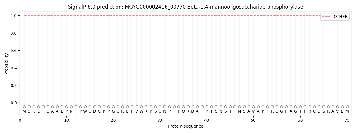
This protein is predicted as OTHER
| Other | SP_Sec_SPI | LIPO_Sec_SPII | TAT_Tat_SPI | TATLIP_Sec_SPII | PILIN_Sec_SPIII |
|---|---|---|---|---|---|
| 1.000041 | 0.000023 | 0.000001 | 0.000000 | 0.000000 | 0.000000 |