You are browsing environment: HUMAN GUT
CAZyme Information: MGYG000002471_01918
You are here: Home > Sequence: MGYG000002471_01918
Basic Information |
Genomic context |
Full Sequence |
Enzyme annotations |
CAZy signature domains |
CDD domains |
CAZyme hits |
PDB hits |
Swiss-Prot hits |
SignalP and Lipop annotations |
TMHMM annotations
Basic Information help
| Species | Yersinia mollaretii | |||||||||||
|---|---|---|---|---|---|---|---|---|---|---|---|---|
| Lineage | Bacteria; Proteobacteria; Gammaproteobacteria; Enterobacterales; Enterobacteriaceae; Yersinia; Yersinia mollaretii | |||||||||||
| CAZyme ID | MGYG000002471_01918 | |||||||||||
| CAZy Family | GH19 | |||||||||||
| CAZyme Description | hypothetical protein | |||||||||||
| CAZyme Property |
|
|||||||||||
| Genome Property |
|
|||||||||||
| Gene Location | Start: 229316; End: 231634 Strand: + | |||||||||||
CDD Domains download full data without filtering help
| Cdd ID | Domain | E-Value | qStart | qEnd | sStart | sEnd | Domain Description |
|---|---|---|---|---|---|---|---|
| pfam11860 | Muraidase | 1.03e-55 | 603 | 768 | 1 | 170 | N-acetylmuramidase. Endolysins are bacteriophage encoded proteins synthesized at the end of the lytic infection cycle. They degrade the peptidoglycan (PG) of the host bacterium to allow viral progeny release. This domain family is found in bacteria and viruses. It is also found associated with pfam01471. One of the family members is the modular Gp110 endolysin found in the Salmonella phage. This domain represents the catalytic region found in the C-terminal of Gp110. It has been demonstrated to have N-acetylmuramidase (lysozyme) activity cleaving the beta-(1,4) glycosidic bond between N-acetylmuramic acid and N-acetylglucosamine residues in the sugar backbone of the PG. Furthermore, sequence alignments containing this domain show that the Gp110 E101 residue is conserved (suggesting that is is the catalytic residue), and followed by serine (a common feature in lysozymes). The structure of endolysins varies depending on their origin. In general, most of the endolysins from phages infecting Gram-positive bacteria have a modular structure consisting of one or two N-terminal enzymatic active domains (EADs) and a C-terminal cell wall binding domain (CBD) separated by a short linker. In silico analysis indicate that this endolysin has a modular structure harboring this EAD family at the C-terminus and a PG_binding_1 CBD at the N-terminus. |
CAZyme Hits help
| Hit ID | E-Value | Query Start | Query End | Hit Start | Hit End |
|---|---|---|---|---|---|
| AHE70536.1 | 3.03e-259 | 1 | 662 | 1 | 671 |
| AWF50744.1 | 3.90e-258 | 1 | 563 | 2 | 564 |
| ANA25340.1 | 6.98e-256 | 1 | 563 | 1 | 563 |
| ANA29683.1 | 6.98e-256 | 1 | 563 | 1 | 563 |
| ANA21118.1 | 6.98e-256 | 1 | 563 | 1 | 563 |
PDB Hits download full data without filtering help
| Hit ID | E-Value | Query Start | Query End | Hit Start | Hit End | Description |
|---|---|---|---|---|---|---|
| 7Q4S_AAA | 1.53e-34 | 588 | 770 | 23 | 204 | ChainAAA, Endolysin [Pseudomonas phage JG004],7Q4S_BBB Chain BBB, Endolysin [Pseudomonas phage JG004],7Q4T_AAA Chain AAA, Endolysin [Pseudomonas phage JG004] |
| 5NM7_A | 7.21e-26 | 588 | 769 | 75 | 259 | Crystalstructure of Burkholderia AP3 phage endolysin [Burkholderia],5NM7_G Crystal structure of Burkholderia AP3 phage endolysin [Burkholderia] |
| 7RUM_A | 4.12e-24 | 586 | 770 | 94 | 283 | ChainA, Endolysin [Salmonella phage GEC_vB_GOT],7RUM_B Chain B, Endolysin [Salmonella phage GEC_vB_GOT] |
Swiss-Prot Hits help
SignalP and Lipop Annotations help
This protein is predicted as OTHER
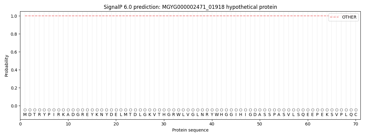
| Other | SP_Sec_SPI | LIPO_Sec_SPII | TAT_Tat_SPI | TATLIP_Sec_SPII | PILIN_Sec_SPIII |
|---|---|---|---|---|---|
| 1.000047 | 0.000000 | 0.000000 | 0.000000 | 0.000000 | 0.000000 |