You are browsing environment: HUMAN GUT
CAZyme Information: MGYG000002471_03770
You are here: Home > Sequence: MGYG000002471_03770
Basic Information |
Genomic context |
Full Sequence |
Enzyme annotations |
CAZy signature domains |
CDD domains |
CAZyme hits |
PDB hits |
Swiss-Prot hits |
SignalP and Lipop annotations |
TMHMM annotations
Basic Information help
| Species | Yersinia mollaretii | |||||||||||
|---|---|---|---|---|---|---|---|---|---|---|---|---|
| Lineage | Bacteria; Proteobacteria; Gammaproteobacteria; Enterobacterales; Enterobacteriaceae; Yersinia; Yersinia mollaretii | |||||||||||
| CAZyme ID | MGYG000002471_03770 | |||||||||||
| CAZy Family | CBM50 | |||||||||||
| CAZyme Description | hypothetical protein | |||||||||||
| CAZyme Property |
|
|||||||||||
| Genome Property |
|
|||||||||||
| Gene Location | Start: 4612; End: 10065 Strand: + | |||||||||||
CDD Domains download full data without filtering help
| Cdd ID | Domain | E-Value | qStart | qEnd | sStart | sEnd | Domain Description |
|---|---|---|---|---|---|---|---|
| cd04059 | Peptidases_S8_Protein_convertases_Kexins_Furin-like | 7.02e-64 | 927 | 1223 | 1 | 297 | Peptidase S8 family domain in Protein convertases. Protein convertases, whose members include furins and kexins, are members of the peptidase S8 or Subtilase clan of proteases. They have an Asp/His/Ser catalytic triad that is not homologous to trypsin. Kexins are involved in the activation of peptide hormones, growth factors, and viral proteins. Furin cleaves cell surface vasoactive peptides and proteins involved in cardiovascular tissue remodeling in the TGN, at cell surface, or in endosomes but rarely in the ER. Furin also plays a key role in blood pressure regulation though the activation of transforming growth factor (TGF)-beta. High specificity is seen for cleavage after dibasic (Lys-Arg or Arg-Arg) or multiple basic residues in protein convertases. There is also strong sequence conservation. |
| cd07498 | Peptidases_S8_15 | 8.92e-29 | 973 | 1218 | 10 | 239 | Peptidase S8 family domain, uncharacterized subfamily 15. This family is a member of the Peptidases S8 or Subtilases serine endo- and exo-peptidase clan. They have an Asp/His/Ser catalytic triad similar to that found in trypsin-like proteases, but do not share their three-dimensional structure and are not homologous to trypsin. The stability of subtilases may be enhanced by calcium, some members have been shown to bind up to 4 ions via binding sites with different affinity. Some members of this clan contain disulfide bonds. These enzymes can be intra- and extracellular, some function at extreme temperatures and pH values. |
| pfam00082 | Peptidase_S8 | 3.37e-27 | 973 | 1254 | 13 | 287 | Subtilase family. Subtilases are a family of serine proteases. They appear to have independently and convergently evolved an Asp/Ser/His catalytic triad, like that found in the trypsin serine proteases (see pfam00089). Structure is an alpha/beta fold containing a 7-stranded parallel beta sheet, order 2314567. |
| cd04848 | Peptidases_S8_Autotransporter_serine_protease_like | 2.36e-23 | 951 | 1223 | 1 | 267 | Peptidase S8 family domain in Autotransporter serine proteases. Autotransporter serine proteases belong to Peptidase S8 or Subtilase family. Subtilases, or subtilisin-like serine proteases, have an Asp/His/Ser catalytic triad similar to that found in trypsin-like proteases, but do not share their three-dimensional structure (an example of convergent evolution). Autotransporters are a superfamily of outer membrane/secreted proteins of gram-negative bacteria. The presence of these subtilisin-like domains in these autotransporters are may enable them to be auto-catalytic and may also serve to allow them to act as a maturation protease cleaving other outer membrane proteins at the cell surface. |
| cd00306 | Peptidases_S8_S53 | 7.75e-22 | 973 | 1221 | 10 | 241 | Peptidase domain in the S8 and S53 families. Members of the peptidases S8 (subtilisin and kexin) and S53 (sedolisin) family include endopeptidases and exopeptidases. The S8 family has an Asp/His/Ser catalytic triad similar to that found in trypsin-like proteases, but do not share their three-dimensional structure and are not homologous to trypsin. Serine acts as a nucleophile, aspartate as an electrophile, and histidine as a base. The S53 family contains a catalytic triad Glu/Asp/Ser with an additional acidic residue Asp in the oxyanion hole, similar to that of subtilisin. The serine residue here is the nucleophilic equivalent of the serine residue in the S8 family, while glutamic acid has the same role here as the histidine base. However, the aspartic acid residue that acts as an electrophile is quite different. In S53, it follows glutamic acid, while in S8 it precedes histidine. The stability of these enzymes may be enhanced by calcium; some members have been shown to bind up to 4 ions via binding sites with different affinity. There is a great diversity in the characteristics of their members: some contain disulfide bonds, some are intracellular while others are extracellular, some function at extreme temperatures, and others at high or low pH values. |
CAZyme Hits help
| Hit ID | E-Value | Query Start | Query End | Hit Start | Hit End |
|---|---|---|---|---|---|
| SBW81129.1 | 2.72e-222 | 264 | 1597 | 309 | 1678 |
| SDU86200.1 | 2.62e-220 | 264 | 1597 | 313 | 1682 |
| AIY26234.1 | 3.76e-09 | 845 | 1223 | 58 | 441 |
| CAG23448.1 | 1.03e-07 | 919 | 1224 | 121 | 443 |
| QZY84418.1 | 1.21e-07 | 870 | 1218 | 95 | 450 |
PDB Hits download full data without filtering help
| Hit ID | E-Value | Query Start | Query End | Hit Start | Hit End | Description |
|---|---|---|---|---|---|---|
| 6YD3_A | 3.50e-33 | 927 | 1390 | 6 | 446 | ChainA, Furin [Homo sapiens],6YD4_A Chain A, Furin [Homo sapiens],6YD7_A Chain A, Furin [Homo sapiens],7O1U_A Chain A, Furin [Homo sapiens],7O1W_A Chain A, Furin [Homo sapiens],7O1Y_A Chain A, Furin [Homo sapiens],7O20_A Chain A, Furin [Homo sapiens],7O22_A Chain A, Furin [Homo sapiens],7QXY_A Chain A, Furin [Homo sapiens],7QXZ_A Chain A, Furin [Homo sapiens],7QY0_A Chain A, Furin [Homo sapiens],7QY1_A Chain A, Furin [Homo sapiens],7QY2_A Chain A, Furin [Homo sapiens] |
| 4OMC_A | 3.61e-33 | 927 | 1390 | 6 | 446 | X-raystructure of human furin in complex with the competitive inhibitor meta-guanidinomethyl-Phac-RVR-Amba [Homo sapiens],4OMC_B X-ray structure of human furin in complex with the competitive inhibitor meta-guanidinomethyl-Phac-RVR-Amba [Homo sapiens],4OMC_C X-ray structure of human furin in complex with the competitive inhibitor meta-guanidinomethyl-Phac-RVR-Amba [Homo sapiens],4OMC_D X-ray structure of human furin in complex with the competitive inhibitor meta-guanidinomethyl-Phac-RVR-Amba [Homo sapiens],4OMC_E X-ray structure of human furin in complex with the competitive inhibitor meta-guanidinomethyl-Phac-RVR-Amba [Homo sapiens],4OMC_F X-ray structure of human furin in complex with the competitive inhibitor meta-guanidinomethyl-Phac-RVR-Amba [Homo sapiens],4OMD_A X-ray structure of human furin in complex with the competitive inhibitor Phac-RVR-Amba [Homo sapiens],4OMD_B X-ray structure of human furin in complex with the competitive inhibitor Phac-RVR-Amba [Homo sapiens],4OMD_C X-ray structure of human furin in complex with the competitive inhibitor Phac-RVR-Amba [Homo sapiens],4OMD_D X-ray structure of human furin in complex with the competitive inhibitor Phac-RVR-Amba [Homo sapiens],4OMD_E X-ray structure of human furin in complex with the competitive inhibitor Phac-RVR-Amba [Homo sapiens],4OMD_F X-ray structure of human furin in complex with the competitive inhibitor Phac-RVR-Amba [Homo sapiens],4RYD_A X-ray structure of human furin in complex with the competitive inhibitor para-guanidinomethyl-Phac-R-Tle-R-Amba [Homo sapiens],4RYD_B X-ray structure of human furin in complex with the competitive inhibitor para-guanidinomethyl-Phac-R-Tle-R-Amba [Homo sapiens],4RYD_C X-ray structure of human furin in complex with the competitive inhibitor para-guanidinomethyl-Phac-R-Tle-R-Amba [Homo sapiens],4RYD_D X-ray structure of human furin in complex with the competitive inhibitor para-guanidinomethyl-Phac-R-Tle-R-Amba [Homo sapiens],4RYD_E X-ray structure of human furin in complex with the competitive inhibitor para-guanidinomethyl-Phac-R-Tle-R-Amba [Homo sapiens],4RYD_F X-ray structure of human furin in complex with the competitive inhibitor para-guanidinomethyl-Phac-R-Tle-R-Amba [Homo sapiens],5JMO_A X-ray structure of furin in complex with the inhibitory antibody Nb14 [Homo sapiens],5JMO_B X-ray structure of furin in complex with the inhibitory antibody Nb14 [Homo sapiens],5JXG_A Structure of the unliganded form of the proprotein convertase furin. [Homo sapiens],5JXH_A Structure the proprotein convertase furin in complex with meta-guanidinomethyl-Phac-RVR-Amba at 2.0 Angstrom resolution. [Homo sapiens],5JXI_A Structure of the unliganded form of the proprotein convertase furin in presence of EDTA. [Homo sapiens],5JXJ_A Structure of the proprotein convertase furin complexed to meta-guanidinomethyl-Phac-RVR-Amba in presence of EDTA [Homo sapiens],5MIM_A Xray structure of human furin bound with the 2,5-dideoxystreptamine derived small molecule inhibitor 1n [Homo sapiens],6EQV_A X-ray structure of the proprotein convertase furin bound with the competitive inhibitor Phac-Cit-Val-Arg-Amba [Homo sapiens],6EQW_A X-ray structure of the proprotein convertase furin bound with the competitive inhibitor 4-aminomethyl-phenylacetyl-Arg-Val-Arg-Amba [Homo sapiens],6EQX_A X-ray structure of the proprotein convertase furin bound with the competitive inhibitor Arg-Arg-Arg-Val-Arg-Amba [Homo sapiens],6HLB_A X-ray structure of furin in complex with the cyclic peptide c[succinyl-Phe-2-Nal-(Arg)4-Lys]-Arg-4-Amba [Homo sapiens],6HLD_A X-ray structure of furin in complex with the cyclic peptide c[succinyl-Phe-2-Nal-(Arg)3-Lys]-Lys-4-Amba [Homo sapiens],6HLE_A X-ray structure of furin in complex with the P6-P2-cyclized peptide H-Lys-Arg-Arg-Tle-Lys-4-Amba [Homo sapiens],6HZA_A X-ray structure of furin in complex with the cyclic peptide c[glutaryl-Arg-Arg-Lys]-Arg-4-Amba [Homo sapiens],6HZB_A X-ray structure of furin in complex with the cyclic inhibitor c[glutaryl-Arg-Arg-Lys]-Lys-4-Amba [Homo sapiens],6HZC_A X-ray structure of furin in complex with the cyclic inhibitor c[glutaryl-BVK-Lys-Arg-Arg-Tle-Lys]-4-Amba [Homo sapiens],6HZD_A X-ray structure of furin in complex with the cyclic inhibitor c[glutaryl-Arg-Arg-Arg-Lys]-Arg-4-Amba [Homo sapiens],6YD2_A Chain A, Furin [Homo sapiens] |
| 4Z2A_A | 1.64e-32 | 927 | 1390 | 4 | 444 | Crystalstructure of unglycosylated apo human furin @1.89A [Homo sapiens] |
| 1P8J_A | 1.80e-32 | 927 | 1390 | 6 | 446 | CrystalStructure Of The Proprotein Convertase Furin [Mus musculus],1P8J_B Crystal Structure Of The Proprotein Convertase Furin [Mus musculus],1P8J_C Crystal Structure Of The Proprotein Convertase Furin [Mus musculus],1P8J_D Crystal Structure Of The Proprotein Convertase Furin [Mus musculus],1P8J_E Crystal Structure Of The Proprotein Convertase Furin [Mus musculus],1P8J_F Crystal Structure Of The Proprotein Convertase Furin [Mus musculus],1P8J_G Crystal Structure Of The Proprotein Convertase Furin [Mus musculus],1P8J_H Crystal Structure Of The Proprotein Convertase Furin [Mus musculus] |
| 7LCU_A | 2.38e-32 | 927 | 1390 | 6 | 446 | ChainA, Furin [Homo sapiens] |
Swiss-Prot Hits download full data without filtering help
| Hit ID | E-Value | Query Start | Query End | Hit Start | Hit End | Description |
|---|---|---|---|---|---|---|
| O17798 | 2.44e-32 | 927 | 1408 | 174 | 642 | Furin-like protease kpc-1 OS=Caenorhabditis elegans OX=6239 GN=kpc-1 PE=1 SV=3 |
| P23377 | 9.69e-32 | 927 | 1390 | 113 | 553 | Furin OS=Rattus norvegicus OX=10116 GN=Furin PE=1 SV=1 |
| Q28193 | 1.30e-31 | 927 | 1390 | 113 | 553 | Furin OS=Bos taurus OX=9913 GN=FURIN PE=1 SV=1 |
| P09958 | 2.96e-31 | 927 | 1390 | 113 | 553 | Furin OS=Homo sapiens OX=9606 GN=FURIN PE=1 SV=2 |
| P23188 | 1.56e-30 | 927 | 1390 | 113 | 553 | Furin OS=Mus musculus OX=10090 GN=Furin PE=1 SV=2 |
SignalP and Lipop Annotations help
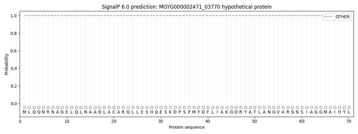
This protein is predicted as OTHER

| Other | SP_Sec_SPI | LIPO_Sec_SPII | TAT_Tat_SPI | TATLIP_Sec_SPII | PILIN_Sec_SPIII |
|---|---|---|---|---|---|
| 1.000042 | 0.000001 | 0.000000 | 0.000000 | 0.000000 | 0.000000 |
