You are browsing environment: HUMAN GUT
CAZyme Information: MGYG000002476_03941
You are here: Home > Sequence: MGYG000002476_03941
Basic Information |
Genomic context |
Full Sequence |
Enzyme annotations |
CAZy signature domains |
CDD domains |
CAZyme hits |
PDB hits |
Swiss-Prot hits |
SignalP and Lipop annotations |
TMHMM annotations
Basic Information help
| Species | Yersinia pestis | |||||||||||
|---|---|---|---|---|---|---|---|---|---|---|---|---|
| Lineage | Bacteria; Proteobacteria; Gammaproteobacteria; Enterobacterales; Enterobacteriaceae; Yersinia; Yersinia pestis | |||||||||||
| CAZyme ID | MGYG000002476_03941 | |||||||||||
| CAZy Family | AA7 | |||||||||||
| CAZyme Description | hypothetical protein | |||||||||||
| CAZyme Property |
|
|||||||||||
| Genome Property |
|
|||||||||||
| Gene Location | Start: 4259133; End: 4261190 Strand: - | |||||||||||
CAZyme Signature Domains help
| Family | Start | End | Evalue | family coverage |
|---|---|---|---|---|
| AA7 | 17 | 535 | 7.2e-164 | 0.9956331877729258 |
CDD Domains download full data without filtering help
| Cdd ID | Domain | E-Value | qStart | qEnd | sStart | sEnd | Domain Description |
|---|---|---|---|---|---|---|---|
| COG3979 | COG3979 | 2.85e-38 | 542 | 685 | 5 | 153 | Chitodextrinase [Carbohydrate transport and metabolism]. |
| COG0277 | GlcD | 4.73e-16 | 18 | 538 | 19 | 459 | FAD/FMN-containing dehydrogenase [Energy production and conversion]. |
| pfam08031 | BBE | 9.10e-11 | 480 | 536 | 1 | 45 | Berberine and berberine like. This domain is found in the berberine bridge and berberine bridge- like enzymes which are involved in the biosynthesis of numerous isoquinoline alkaloids. They catalyze the transformation of the N-methyl group of (S)-reticuline into the C-8 berberine bridge carbon of (S)-scoulerine. |
| cd00063 | FN3 | 5.81e-08 | 542 | 628 | 1 | 93 | Fibronectin type 3 domain; One of three types of internal repeats found in the plasma protein fibronectin. Its tenth fibronectin type III repeat contains an RGD cell recognition sequence in a flexible loop between 2 strands. Approximately 2% of all animal proteins contain the FN3 repeat; including extracellular and intracellular proteins, membrane spanning cytokine receptors, growth hormone receptors, tyrosine phosphatase receptors, and adhesion molecules. FN3-like domains are also found in bacterial glycosyl hydrolases. |
| cd12214 | ChiA1_BD | 1.12e-06 | 634 | 678 | 1 | 42 | chitin-binding domain of Chi A1-like proteins. This group contains proteins related to the chitin binding domain of chitinase A1 (ChiA1) of Bacillus circulans WL-12. Glycosidase ChiA1 hydrolyzes chitin and is comprised of several domains: the C-terminal chitin binding domain, an N-terminal and catalytic domain, and 2 fibronectin type III-like domains. Chitinases function in invertebrates in the degradation of old exoskeletons, in fungi to utilize chitin in cell walls, and in bacteria which use chitin as an energy source. Bacillus circulans WL-12 ChiA1 facilitates invasion of fungal cell walls. The ChiAi chitin binding domain is required for the specific recognition of insoluble chitin. although topologically and structurally related, ChiA1 lacks the characteristic aromatic residues of Erwinia chrysanthemi endoglucanase Z (CBD(EGZ)). |
CAZyme Hits help
| Hit ID | E-Value | Query Start | Query End | Hit Start | Hit End |
|---|---|---|---|---|---|
| AYX00436.1 | 0.0 | 1 | 685 | 1 | 685 |
| VEE73860.1 | 0.0 | 1 | 685 | 1 | 685 |
| AZA32001.1 | 0.0 | 1 | 685 | 1 | 685 |
| AYW95221.1 | 0.0 | 1 | 685 | 1 | 685 |
| AYW85797.1 | 0.0 | 1 | 685 | 1 | 685 |
PDB Hits download full data without filtering help
| Hit ID | E-Value | Query Start | Query End | Hit Start | Hit End | Description |
|---|---|---|---|---|---|---|
| 2Y08_A | 2.55e-66 | 11 | 535 | 44 | 525 | Structureof the substrate-free FAD-dependent tirandamycin oxidase TamL [Streptomyces sp. 307-9],2Y08_B Structure of the substrate-free FAD-dependent tirandamycin oxidase TamL [Streptomyces sp. 307-9],2Y3R_A Structure of the tirandamycin-bound FAD-dependent tirandamycin oxidase TamL in P21 space group [Streptomyces sp. 307-9],2Y3R_B Structure of the tirandamycin-bound FAD-dependent tirandamycin oxidase TamL in P21 space group [Streptomyces sp. 307-9],2Y3R_C Structure of the tirandamycin-bound FAD-dependent tirandamycin oxidase TamL in P21 space group [Streptomyces sp. 307-9],2Y3R_D Structure of the tirandamycin-bound FAD-dependent tirandamycin oxidase TamL in P21 space group [Streptomyces sp. 307-9],2Y3S_A Structure of the tirandamycine-bound FAD-dependent tirandamycin oxidase TamL in C2 space group [Streptomyces sp. 307-9],2Y3S_B Structure of the tirandamycine-bound FAD-dependent tirandamycin oxidase TamL in C2 space group [Streptomyces sp. 307-9],2Y4G_A Structure of the Tirandamycin-bound FAD-dependent tirandamycin oxidase TamL in P212121 space group [Streptomyces sp. 307-9],2Y4G_B Structure of the Tirandamycin-bound FAD-dependent tirandamycin oxidase TamL in P212121 space group [Streptomyces sp. 307-9] |
| 2IPI_A | 7.68e-64 | 4 | 535 | 33 | 517 | CrystalStructure of Aclacinomycin Oxidoreductase [Streptomyces galilaeus],2IPI_B Crystal Structure of Aclacinomycin Oxidoreductase [Streptomyces galilaeus],2IPI_C Crystal Structure of Aclacinomycin Oxidoreductase [Streptomyces galilaeus],2IPI_D Crystal Structure of Aclacinomycin Oxidoreductase [Streptomyces galilaeus] |
| 5I1V_A | 1.19e-58 | 41 | 535 | 42 | 497 | Crystalstructure of CrmK, a flavoenzyme involved in the shunt product recycling mechanism in caerulomycin biosynthesis [Actinoalloteichus sp. WH1-2216-6],5I1V_B Crystal structure of CrmK, a flavoenzyme involved in the shunt product recycling mechanism in caerulomycin biosynthesis [Actinoalloteichus sp. WH1-2216-6],5I1V_C Crystal structure of CrmK, a flavoenzyme involved in the shunt product recycling mechanism in caerulomycin biosynthesis [Actinoalloteichus sp. WH1-2216-6],5I1V_D Crystal structure of CrmK, a flavoenzyme involved in the shunt product recycling mechanism in caerulomycin biosynthesis [Actinoalloteichus sp. WH1-2216-6],5I1W_A Crystal structure of CrmK, a flavoenzyme involved in the shunt product recycling mechanism in caerulomycin biosynthesis [Actinoalloteichus sp. WH1-2216-6],5I1W_B Crystal structure of CrmK, a flavoenzyme involved in the shunt product recycling mechanism in caerulomycin biosynthesis [Actinoalloteichus sp. WH1-2216-6],5I1W_C Crystal structure of CrmK, a flavoenzyme involved in the shunt product recycling mechanism in caerulomycin biosynthesis [Actinoalloteichus sp. WH1-2216-6],5I1W_D Crystal structure of CrmK, a flavoenzyme involved in the shunt product recycling mechanism in caerulomycin biosynthesis [Actinoalloteichus sp. WH1-2216-6] |
| 2WDW_A | 5.97e-54 | 33 | 535 | 61 | 521 | TheNative Crystal Structure of the Primary Hexose Oxidase (Dbv29) in Antibiotic A40926 Biosynthesis [Nonomuraea gerenzanensis],2WDW_B The Native Crystal Structure of the Primary Hexose Oxidase (Dbv29) in Antibiotic A40926 Biosynthesis [Nonomuraea gerenzanensis],5AWV_A Crystal structure of glycopeptide hexose oxidase DBV29 complexed with teicoplanin [Nonomuraea gerenzanensis],5AWV_B Crystal structure of glycopeptide hexose oxidase DBV29 complexed with teicoplanin [Nonomuraea gerenzanensis],5AWV_C Crystal structure of glycopeptide hexose oxidase DBV29 complexed with teicoplanin [Nonomuraea gerenzanensis],5AWV_D Crystal structure of glycopeptide hexose oxidase DBV29 complexed with teicoplanin [Nonomuraea gerenzanensis] |
| 3POP_A | 6.54e-52 | 31 | 537 | 36 | 501 | Thecrystal structure of GilR, an oxidoreductase that catalyzes the terminal step of gilvocarcin biosynthesis [Streptomyces griseoflavus],3POP_B The crystal structure of GilR, an oxidoreductase that catalyzes the terminal step of gilvocarcin biosynthesis [Streptomyces griseoflavus],3POP_C The crystal structure of GilR, an oxidoreductase that catalyzes the terminal step of gilvocarcin biosynthesis [Streptomyces griseoflavus],3POP_D The crystal structure of GilR, an oxidoreductase that catalyzes the terminal step of gilvocarcin biosynthesis [Streptomyces griseoflavus],3PQB_A The crystal structure of pregilvocarcin in complex with GilR, an oxidoreductase that catalyzes the terminal step of gilvocarcin biosynthesis [Streptomyces griseoflavus],3PQB_B The crystal structure of pregilvocarcin in complex with GilR, an oxidoreductase that catalyzes the terminal step of gilvocarcin biosynthesis [Streptomyces griseoflavus],3PQB_C The crystal structure of pregilvocarcin in complex with GilR, an oxidoreductase that catalyzes the terminal step of gilvocarcin biosynthesis [Streptomyces griseoflavus],3PQB_D The crystal structure of pregilvocarcin in complex with GilR, an oxidoreductase that catalyzes the terminal step of gilvocarcin biosynthesis [Streptomyces griseoflavus] |
Swiss-Prot Hits download full data without filtering help
| Hit ID | E-Value | Query Start | Query End | Hit Start | Hit End | Description |
|---|---|---|---|---|---|---|
| A3RXB7 | 1.62e-122 | 11 | 535 | 12 | 506 | N-acetyl-D-hexosamine oxidase OS=Ralstonia solanacearum (strain UW551) OX=342110 GN=RRSL_02030 PE=1 SV=1 |
| P93762 | 1.41e-81 | 3 | 536 | 13 | 535 | Hexose oxidase OS=Chondrus crispus OX=2769 GN=HOX PE=1 SV=2 |
| Q0PCD7 | 7.14e-63 | 4 | 535 | 57 | 541 | Aclacinomycin-N/aclacinomycin-A oxidase OS=Streptomyces galilaeus OX=33899 GN=aknOx PE=1 SV=1 |
| Q796Y5 | 1.61e-27 | 2 | 536 | 9 | 443 | Uncharacterized FAD-linked oxidoreductase YgaK OS=Bacillus subtilis (strain 168) OX=224308 GN=ygaK PE=3 SV=4 |
| Q5QJ60 | 1.47e-25 | 27 | 536 | 75 | 519 | Berberine bridge enzyme-like Cyn d 4 OS=Cynodon dactylon OX=28909 PE=1 SV=1 |
SignalP and Lipop Annotations help
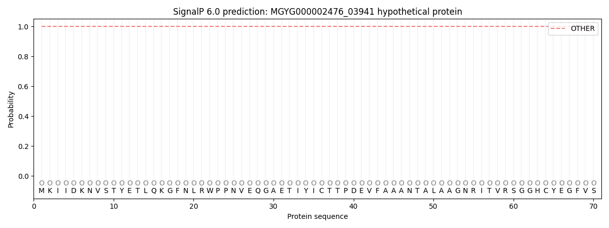
This protein is predicted as OTHER
| Other | SP_Sec_SPI | LIPO_Sec_SPII | TAT_Tat_SPI | TATLIP_Sec_SPII | PILIN_Sec_SPIII |
|---|---|---|---|---|---|
| 1.000043 | 0.000000 | 0.000000 | 0.000000 | 0.000000 | 0.000000 |