You are browsing environment: HUMAN GUT
CAZyme Information: MGYG000002525_02497
You are here: Home > Sequence: MGYG000002525_02497
Basic Information |
Genomic context |
Full Sequence |
Enzyme annotations |
CAZy signature domains |
CDD domains |
CAZyme hits |
PDB hits |
Swiss-Prot hits |
SignalP and Lipop annotations |
TMHMM annotations
Basic Information help
| Species | Yersinia intermedia | |||||||||||
|---|---|---|---|---|---|---|---|---|---|---|---|---|
| Lineage | Bacteria; Proteobacteria; Gammaproteobacteria; Enterobacterales; Enterobacteriaceae; Yersinia; Yersinia intermedia | |||||||||||
| CAZyme ID | MGYG000002525_02497 | |||||||||||
| CAZy Family | GT41 | |||||||||||
| CAZyme Description | UDP-glucose:protein N-beta-glucosyltransferase | |||||||||||
| CAZyme Property |
|
|||||||||||
| Genome Property |
|
|||||||||||
| Gene Location | Start: 34650; End: 36500 Strand: + | |||||||||||
CAZyme Signature Domains help
| Family | Start | End | Evalue | family coverage |
|---|---|---|---|---|
| GT41 | 52 | 594 | 3.5e-44 | 0.47092198581560285 |
CDD Domains download full data without filtering help
| Cdd ID | Domain | E-Value | qStart | qEnd | sStart | sEnd | Domain Description |
|---|---|---|---|---|---|---|---|
| pfam18071 | HMW1C_N | 4.72e-35 | 18 | 153 | 2 | 143 | HMW1C N-terminal. This is the N-terminal domain found in Actinobacillus pleuropneumoniae HMW1C (ApHMW1C). HMW1 adhesin is an N-linked glycoprotein that mediates adherence to respiratory epithelium through N-glycosylation of protein acceptor sites an O-glycosylation of sugar acceptor sites. The N-terminal domain forms an all alpha domain (AAD) when combined with the domain spanning from residue 154 to residue 245. The AAD interacts extensively with the C-terminal GT-B fold in order to create unique groove with potential to accommodate the acceptor protein. |
| 408070 | pfam18254 | 2.46e-25 | 158 | 245 | 1 | 88 | HMw1_D2 HMW1 domain 2. This domain is found in Actinobacillus pleuropneumoniae HMW1C (ApHMW1C). HMW1 adhesin is an N-linked glycoprotein that mediates adherence to respiratory epithelium through N-glycosylation of protein acceptor sites and O-glycosylation of sugar acceptor sites. This domain forms an all alpha domain (AAD) when combined with the N-terminal domain. The AAD interacts extensively with the C-terminal GT-B fold in order to create a unique groove with the potential to accommodate the acceptor protein. |
| COG3914 | Spy | 2.33e-12 | 356 | 597 | 356 | 601 | Predicted O-linked N-acetylglucosamine transferase, SPINDLY family [Posttranslational modification, protein turnover, chaperones]. |
| pfam01568 | Molydop_binding | 0.003 | 271 | 342 | 2 | 77 | Molydopterin dinucleotide binding domain. This domain is found in various molybdopterin - containing oxidoreductases and tungsten formylmethanofuran dehydrogenase subunit d (FwdD) and molybdenum formylmethanofuran dehydrogenase subunit (FmdD); where the domain constitutes almost the entire subunit. The formylmethanofuran dehydrogenase catalyzes the first step in methane formation from CO2 in methanogenic archaea and has a molybdopterin dinucleotide cofactor. This domain corresponds to the C-terminal domain IV in dimethyl sulfoxide (DMSO)reductase which interacts with the 2-amino pyrimidone ring of both molybdopterin guanine dinucleotide molecules. |
CAZyme Hits help
| Hit ID | E-Value | Query Start | Query End | Hit Start | Hit End |
|---|---|---|---|---|---|
| AVL36032.1 | 0.0 | 1 | 616 | 1 | 616 |
| ARB86181.1 | 0.0 | 1 | 616 | 1 | 616 |
| VDZ58263.1 | 0.0 | 1 | 616 | 1 | 616 |
| QGR65326.1 | 0.0 | 1 | 616 | 1 | 616 |
| QGR70343.1 | 0.0 | 1 | 616 | 1 | 616 |
PDB Hits download full data without filtering help
| Hit ID | E-Value | Query Start | Query End | Hit Start | Hit End | Description |
|---|---|---|---|---|---|---|
| 3Q3E_A | 2.27e-147 | 23 | 604 | 23 | 619 | Crystalstructure of the Actinobacillus pleuropneumoniae HMW1C glycosyltransferase [Actinobacillus pleuropneumoniae serovar 1 str. 4074],3Q3E_B Crystal structure of the Actinobacillus pleuropneumoniae HMW1C glycosyltransferase [Actinobacillus pleuropneumoniae serovar 1 str. 4074],3Q3H_A Crystal structure of the Actinobacillus pleuropneumoniae HMW1C glycosyltransferase in complex with UDP-GLC [Actinobacillus pleuropneumoniae serovar 1 str. 4074],3Q3H_B Crystal structure of the Actinobacillus pleuropneumoniae HMW1C glycosyltransferase in complex with UDP-GLC [Actinobacillus pleuropneumoniae serovar 1 str. 4074],3Q3I_A Crystal structure of the Actinobacillus pleuropneumoniae HMW1C glycosyltransferase in the presence of peptide N1131 [Actinobacillus pleuropneumoniae serovar 1 str. 4074],3Q3I_B Crystal structure of the Actinobacillus pleuropneumoniae HMW1C glycosyltransferase in the presence of peptide N1131 [Actinobacillus pleuropneumoniae serovar 1 str. 4074] |
Swiss-Prot Hits download full data without filtering help
| Hit ID | E-Value | Query Start | Query End | Hit Start | Hit End | Description |
|---|---|---|---|---|---|---|
| A3N2T3 | 1.77e-146 | 23 | 604 | 12 | 608 | UDP-glucose:protein N-beta-glucosyltransferase OS=Actinobacillus pleuropneumoniae serotype 5b (strain L20) OX=416269 GN=APL_1635 PE=1 SV=1 |
| B3H2N2 | 3.10e-138 | 23 | 604 | 12 | 608 | UDP-glucose:protein N-beta-glucosyltransferase OS=Actinobacillus pleuropneumoniae serotype 7 (strain AP76) OX=537457 GN=APP7_1697 PE=1 SV=1 |
SignalP and Lipop Annotations help
This protein is predicted as OTHER
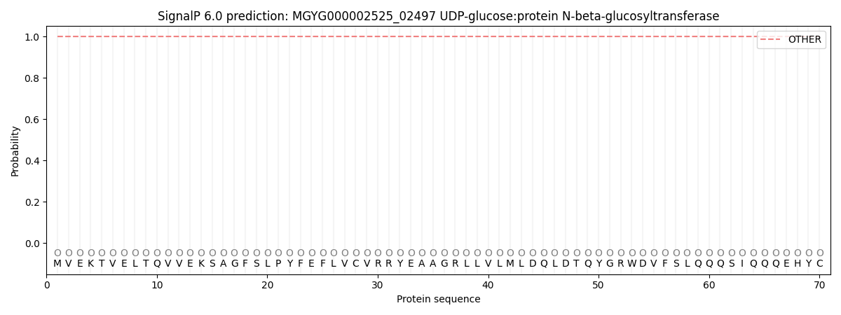
| Other | SP_Sec_SPI | LIPO_Sec_SPII | TAT_Tat_SPI | TATLIP_Sec_SPII | PILIN_Sec_SPIII |
|---|---|---|---|---|---|
| 1.000052 | 0.000000 | 0.000000 | 0.000000 | 0.000000 | 0.000000 |