You are browsing environment: HUMAN GUT
CAZyme Information: MGYG000002554_00399
You are here: Home > Sequence: MGYG000002554_00399
Basic Information |
Genomic context |
Full Sequence |
Enzyme annotations |
CAZy signature domains |
CDD domains |
CAZyme hits |
PDB hits |
Swiss-Prot hits |
SignalP and Lipop annotations |
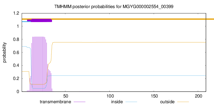
TMHMM annotations
Basic Information help
| Species | Streptococcus oralis_W | |||||||||||
|---|---|---|---|---|---|---|---|---|---|---|---|---|
| Lineage | Bacteria; Firmicutes; Bacilli; Lactobacillales; Streptococcaceae; Streptococcus; Streptococcus oralis_W | |||||||||||
| CAZyme ID | MGYG000002554_00399 | |||||||||||
| CAZy Family | CBM50 | |||||||||||
| CAZyme Description | hypothetical protein | |||||||||||
| CAZyme Property |
|
|||||||||||
| Genome Property |
|
|||||||||||
| Gene Location | Start: 18; End: 647 Strand: - | |||||||||||
CDD Domains download full data without filtering help
| Cdd ID | Domain | E-Value | qStart | qEnd | sStart | sEnd | Domain Description |
|---|---|---|---|---|---|---|---|
| cd00118 | LysM | 1.83e-13 | 33 | 77 | 1 | 45 | Lysin Motif is a small domain involved in binding peptidoglycan. LysM, a small globular domain with approximately 40 amino acids, is a widespread protein module involved in binding peptidoglycan in bacteria and chitin in eukaryotes. The domain was originally identified in enzymes that degrade bacterial cell walls, but proteins involved in many other biological functions also contain this domain. It has been reported that the LysM domain functions as a signal for specific plant-bacteria recognition in bacterial pathogenesis. Many of these enzymes are modular and are composed of catalytic units linked to one or several repeats of LysM domains. LysM domains are found in bacteria and eukaryotes. |
| smart00257 | LysM | 2.45e-13 | 34 | 77 | 1 | 44 | Lysin motif. |
| pfam01476 | LysM | 5.81e-13 | 35 | 77 | 1 | 42 | LysM domain. The LysM (lysin motif) domain is about 40 residues long. It is found in a variety of enzymes involved in bacterial cell wall degradation. This domain may have a general peptidoglycan binding function. The structure of this domain is known. |
| PRK11198 | PRK11198 | 9.12e-06 | 29 | 77 | 92 | 145 | LysM domain/BON superfamily protein; Provisional |
| COG1388 | LysM | 1.37e-05 | 28 | 77 | 62 | 110 | LysM repeat [Cell wall/membrane/envelope biogenesis]. |
CAZyme Hits help
| Hit ID | E-Value | Query Start | Query End | Hit Start | Hit End |
|---|---|---|---|---|---|
| AQA07376.1 | 1.61e-112 | 1 | 209 | 1 | 206 |
| BAV80643.1 | 2.15e-111 | 1 | 209 | 1 | 210 |
| CBZ01503.1 | 3.86e-109 | 1 | 209 | 1 | 208 |
| VEF79810.1 | 3.35e-106 | 1 | 209 | 1 | 211 |
| QQL00262.1 | 1.93e-105 | 1 | 209 | 1 | 211 |
Swiss-Prot Hits download full data without filtering help
| Hit ID | E-Value | Query Start | Query End | Hit Start | Hit End | Description |
|---|---|---|---|---|---|---|
| P54421 | 1.73e-07 | 34 | 85 | 150 | 200 | Probable peptidoglycan endopeptidase LytE OS=Bacillus subtilis (strain 168) OX=224308 GN=lytE PE=1 SV=1 |
| O07532 | 2.29e-06 | 34 | 84 | 308 | 357 | Peptidoglycan endopeptidase LytF OS=Bacillus subtilis (strain 168) OX=224308 GN=lytF PE=1 SV=2 |
| O34669 | 7.69e-06 | 28 | 89 | 70 | 132 | Cell wall-binding protein YocH OS=Bacillus subtilis (strain 168) OX=224308 GN=yocH PE=1 SV=1 |
| Q49UX4 | 8.48e-06 | 34 | 85 | 87 | 137 | N-acetylmuramoyl-L-alanine amidase sle1 OS=Staphylococcus saprophyticus subsp. saprophyticus (strain ATCC 15305 / DSM 20229 / NCIMB 8711 / NCTC 7292 / S-41) OX=342451 GN=sle1 PE=3 SV=1 |
SignalP and Lipop Annotations help
This protein is predicted as SP
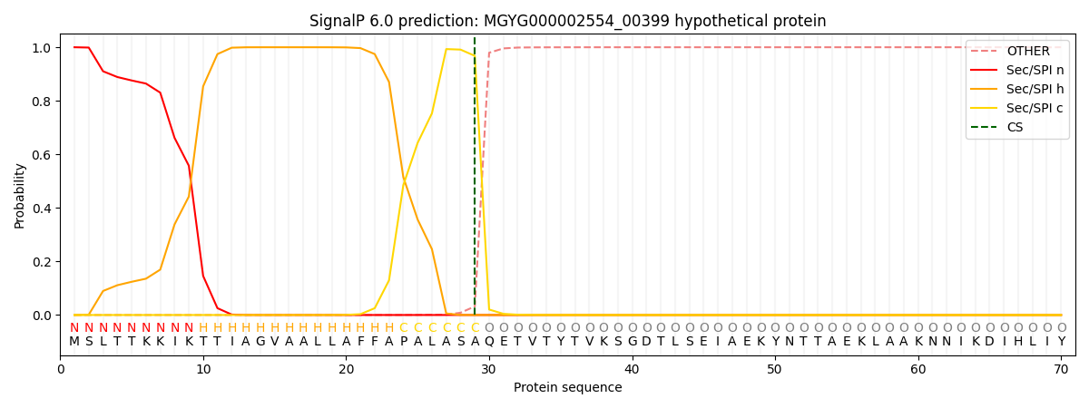
| Other | SP_Sec_SPI | LIPO_Sec_SPII | TAT_Tat_SPI | TATLIP_Sec_SPII | PILIN_Sec_SPIII |
|---|---|---|---|---|---|
| 0.000405 | 0.998755 | 0.000207 | 0.000222 | 0.000203 | 0.000178 |