You are browsing environment: HUMAN GUT
CAZyme Information: MGYG000002895_00874
You are here: Home > Sequence: MGYG000002895_00874
Basic Information |
Genomic context |
Full Sequence |
Enzyme annotations |
CAZy signature domains |
CDD domains |
CAZyme hits |
PDB hits |
Swiss-Prot hits |
SignalP and Lipop annotations |
TMHMM annotations
Basic Information help
| Species | Lawsonella clevelandensis | |||||||||||
|---|---|---|---|---|---|---|---|---|---|---|---|---|
| Lineage | Bacteria; Actinobacteriota; Actinomycetia; Mycobacteriales; Mycobacteriaceae; Lawsonella; Lawsonella clevelandensis | |||||||||||
| CAZyme ID | MGYG000002895_00874 | |||||||||||
| CAZy Family | GT4 | |||||||||||
| CAZyme Description | D-inositol 3-phosphate glycosyltransferase | |||||||||||
| CAZyme Property |
|
|||||||||||
| Genome Property |
|
|||||||||||
| Gene Location | Start: 4883; End: 5410 Strand: - | |||||||||||
CDD Domains download full data without filtering help
| Cdd ID | Domain | E-Value | qStart | qEnd | sStart | sEnd | Domain Description |
|---|---|---|---|---|---|---|---|
| TIGR03449 | mycothiol_MshA | 4.23e-82 | 2 | 167 | 237 | 402 | D-inositol-3-phosphate glycosyltransferase. Members of this protein family, found exclusively in the Actinobacteria, are MshA, the glycosyltransferase of mycothiol biosynthesis. Mycothiol replaces glutathione in these species. |
| cd03800 | GT4_sucrose_synthase | 3.65e-48 | 1 | 162 | 237 | 398 | sucrose-phosphate synthase and similar proteins. This family is most closely related to the GT4 family of glycosyltransferases. The sucrose-phosphate synthases in this family may be unique to plants and photosynthetic bacteria. This enzyme catalyzes the synthesis of sucrose 6-phosphate from fructose 6-phosphate and uridine 5'-diphosphate-glucose, a key regulatory step of sucrose metabolism. The activity of this enzyme is regulated by phosphorylation and moderated by the concentration of various metabolites and light. |
| COG0438 | RfaB | 7.25e-38 | 1 | 168 | 216 | 378 | Glycosyltransferase involved in cell wall bisynthesis [Cell wall/membrane/envelope biogenesis]. |
| pfam00534 | Glycos_transf_1 | 1.80e-35 | 1 | 137 | 19 | 148 | Glycosyl transferases group 1. Mutations in this domain of PIGA lead to disease (Paroxysmal Nocturnal haemoglobinuria). Members of this family transfer activated sugars to a variety of substrates, including glycogen, Fructose-6-phosphate and lipopolysaccharides. Members of this family transfer UDP, ADP, GDP or CMP linked sugars. The eukaryotic glycogen synthases may be distant members of this family. |
| cd03801 | GT4_PimA-like | 4.96e-35 | 2 | 165 | 210 | 366 | phosphatidyl-myo-inositol mannosyltransferase. This family is most closely related to the GT4 family of glycosyltransferases and named after PimA in Propionibacterium freudenreichii, which is involved in the biosynthesis of phosphatidyl-myo-inositol mannosides (PIM) which are early precursors in the biosynthesis of lipomannans (LM) and lipoarabinomannans (LAM), and catalyzes the addition of a mannosyl residue from GDP-D-mannose (GDP-Man) to the position 2 of the carrier lipid phosphatidyl-myo-inositol (PI) to generate a phosphatidyl-myo-inositol bearing an alpha-1,2-linked mannose residue (PIM1). Glycosyltransferases catalyze the transfer of sugar moieties from activated donor molecules to specific acceptor molecules, forming glycosidic bonds. The acceptor molecule can be a lipid, a protein, a heterocyclic compound, or another carbohydrate residue. This group of glycosyltransferases is most closely related to the previously defined glycosyltransferase family 1 (GT1). The members of this family may transfer UDP, ADP, GDP, or CMP linked sugars. The diverse enzymatic activities among members of this family reflect a wide range of biological functions. The protein structure available for this family has the GTB topology, one of the two protein topologies observed for nucleotide-sugar-dependent glycosyltransferases. GTB proteins have distinct N- and C- terminal domains each containing a typical Rossmann fold. The two domains have high structural homology despite minimal sequence homology. The large cleft that separates the two domains includes the catalytic center and permits a high degree of flexibility. The members of this family are found mainly in certain bacteria and archaea. |
CAZyme Hits help
| Hit ID | E-Value | Query Start | Query End | Hit Start | Hit End |
|---|---|---|---|---|---|
| VHO00307.1 | 1.95e-119 | 1 | 175 | 266 | 440 |
| ALE18849.1 | 5.73e-119 | 1 | 175 | 256 | 430 |
| APT89913.1 | 5.64e-63 | 1 | 167 | 249 | 415 |
| AGP31678.1 | 9.95e-62 | 1 | 167 | 252 | 418 |
| AWH93987.1 | 1.31e-61 | 1 | 164 | 256 | 419 |
PDB Hits download full data without filtering help
| Hit ID | E-Value | Query Start | Query End | Hit Start | Hit End | Description |
|---|---|---|---|---|---|---|
| 3C4Q_A | 1.27e-53 | 1 | 170 | 240 | 408 | Structureof the retaining glycosyltransferase MshA : The first step in mycothiol biosynthesis. Organism : Corynebacterium glutamicum- Complex with UDP [Corynebacterium glutamicum],3C4Q_B Structure of the retaining glycosyltransferase MshA : The first step in mycothiol biosynthesis. Organism : Corynebacterium glutamicum- Complex with UDP [Corynebacterium glutamicum],3C4V_A Structure of the retaining glycosyltransferase MshA:The first step in mycothiol biosynthesis. Organism: Corynebacterium glutamicum : Complex with UDP and 1L-INS-1-P. [Corynebacterium glutamicum],3C4V_B Structure of the retaining glycosyltransferase MshA:The first step in mycothiol biosynthesis. Organism: Corynebacterium glutamicum : Complex with UDP and 1L-INS-1-P. [Corynebacterium glutamicum] |
| 3C48_A | 1.64e-53 | 1 | 170 | 260 | 428 | Structureof the retaining glycosyltransferase MshA: The first step in mycothiol biosynthesis. Organism: Corynebacterium glutamicum- APO (OPEN) structure. [Corynebacterium glutamicum],3C48_B Structure of the retaining glycosyltransferase MshA: The first step in mycothiol biosynthesis. Organism: Corynebacterium glutamicum- APO (OPEN) structure. [Corynebacterium glutamicum] |
| 4N9W_A | 2.99e-16 | 1 | 164 | 210 | 364 | Crystalstructure of phosphatidyl mannosyltransferase PimA [Mycolicibacterium smegmatis MC2 155],4NC9_A Crystal structure of phosphatidyl mannosyltransferase PimA [Mycolicibacterium smegmatis MC2 155],4NC9_B Crystal structure of phosphatidyl mannosyltransferase PimA [Mycolicibacterium smegmatis MC2 155],4NC9_C Crystal structure of phosphatidyl mannosyltransferase PimA [Mycolicibacterium smegmatis MC2 155],4NC9_D Crystal structure of phosphatidyl mannosyltransferase PimA [Mycolicibacterium smegmatis MC2 155] |
| 2GEJ_A | 3.16e-16 | 1 | 164 | 226 | 380 | CrystalStructure of phosphatidylinositol mannosyltransferase (PimA) from Mycobacterium smegmatis in complex with GDP-Man [Mycolicibacterium smegmatis MC2 155],2GEK_A Crystal Structure of phosphatidylinositol mannosyltransferase (PimA) from Mycobacterium smegmatis in complex with GDP [Mycolicibacterium smegmatis MC2 155] |
| 6KIH_A | 3.08e-15 | 3 | 166 | 259 | 423 | Sucrose-phosphatesynthase (tll1590) from Thermosynechococcus elongatus [Thermosynechococcus vestitus],6KIH_B Sucrose-phosphate synthase (tll1590) from Thermosynechococcus elongatus [Thermosynechococcus vestitus],6KIH_C Sucrose-phosphate synthase (tll1590) from Thermosynechococcus elongatus [Thermosynechococcus vestitus],6KIH_D Sucrose-phosphate synthase (tll1590) from Thermosynechococcus elongatus [Thermosynechococcus vestitus],6KIH_E Sucrose-phosphate synthase (tll1590) from Thermosynechococcus elongatus [Thermosynechococcus vestitus],6KIH_F Sucrose-phosphate synthase (tll1590) from Thermosynechococcus elongatus [Thermosynechococcus vestitus],6KIH_G Sucrose-phosphate synthase (tll1590) from Thermosynechococcus elongatus [Thermosynechococcus vestitus],6KIH_H Sucrose-phosphate synthase (tll1590) from Thermosynechococcus elongatus [Thermosynechococcus vestitus],6KIH_I Sucrose-phosphate synthase (tll1590) from Thermosynechococcus elongatus [Thermosynechococcus vestitus],6KIH_J Sucrose-phosphate synthase (tll1590) from Thermosynechococcus elongatus [Thermosynechococcus vestitus],6KIH_K Sucrose-phosphate synthase (tll1590) from Thermosynechococcus elongatus [Thermosynechococcus vestitus],6KIH_L Sucrose-phosphate synthase (tll1590) from Thermosynechococcus elongatus [Thermosynechococcus vestitus] |
Swiss-Prot Hits download full data without filtering help
| Hit ID | E-Value | Query Start | Query End | Hit Start | Hit End | Description |
|---|---|---|---|---|---|---|
| B1VEI4 | 6.09e-61 | 1 | 165 | 240 | 404 | D-inositol 3-phosphate glycosyltransferase OS=Corynebacterium urealyticum (strain ATCC 43042 / DSM 7109) OX=504474 GN=mshA PE=3 SV=1 |
| Q4JSW2 | 4.71e-59 | 1 | 164 | 240 | 403 | D-inositol 3-phosphate glycosyltransferase OS=Corynebacterium jeikeium (strain K411) OX=306537 GN=mshA PE=3 SV=1 |
| Q0SF06 | 8.58e-57 | 1 | 171 | 254 | 424 | D-inositol 3-phosphate glycosyltransferase OS=Rhodococcus jostii (strain RHA1) OX=101510 GN=mshA PE=3 SV=1 |
| Q5YP47 | 2.34e-56 | 1 | 167 | 250 | 416 | D-inositol 3-phosphate glycosyltransferase OS=Nocardia farcinica (strain IFM 10152) OX=247156 GN=mshA PE=3 SV=1 |
| Q8FSH1 | 2.54e-56 | 1 | 167 | 240 | 405 | D-inositol 3-phosphate glycosyltransferase OS=Corynebacterium efficiens (strain DSM 44549 / YS-314 / AJ 12310 / JCM 11189 / NBRC 100395) OX=196164 GN=mshA PE=3 SV=1 |
SignalP and Lipop Annotations help
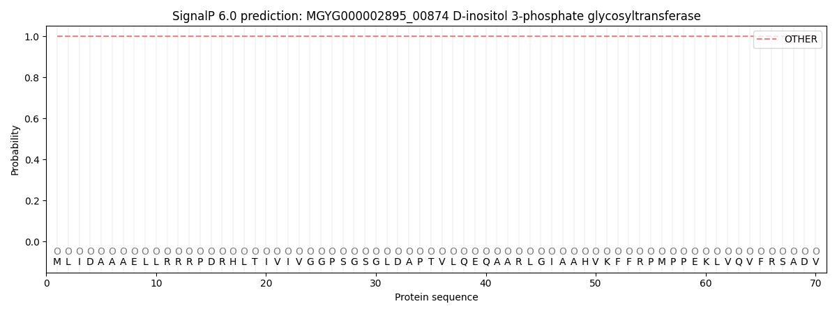
This protein is predicted as OTHER
| Other | SP_Sec_SPI | LIPO_Sec_SPII | TAT_Tat_SPI | TATLIP_Sec_SPII | PILIN_Sec_SPIII |
|---|---|---|---|---|---|
| 1.000030 | 0.000000 | 0.000000 | 0.000000 | 0.000000 | 0.000000 |