You are browsing environment: HUMAN GUT
CAZyme Information: MGYG000003137_07423
You are here: Home > Sequence: MGYG000003137_07423
Basic Information |
Genomic context |
Full Sequence |
Enzyme annotations |
CAZy signature domains |
CDD domains |
CAZyme hits |
PDB hits |
Swiss-Prot hits |
SignalP and Lipop annotations |
TMHMM annotations
Basic Information help
| Species | Bradyrhizobium sp000015165 | |||||||||||
|---|---|---|---|---|---|---|---|---|---|---|---|---|
| Lineage | Bacteria; Proteobacteria; Alphaproteobacteria; Rhizobiales; Xanthobacteraceae; Bradyrhizobium; Bradyrhizobium sp000015165 | |||||||||||
| CAZyme ID | MGYG000003137_07423 | |||||||||||
| CAZy Family | GH13 | |||||||||||
| CAZyme Description | 1,4-alpha-glucan branching enzyme GlgB | |||||||||||
| CAZyme Property |
|
|||||||||||
| Genome Property |
|
|||||||||||
| Gene Location | Start: 13395; End: 15356 Strand: - | |||||||||||
CAZyme Signature Domains help
| Family | Start | End | Evalue | family coverage |
|---|---|---|---|---|
| GH13 | 155 | 500 | 1.2e-37 | 0.9498327759197325 |
CDD Domains download full data without filtering help
| Cdd ID | Domain | E-Value | qStart | qEnd | sStart | sEnd | Domain Description |
|---|---|---|---|---|---|---|---|
| cd11325 | AmyAc_GTHase | 1.22e-134 | 105 | 570 | 1 | 436 | Alpha amylase catalytic domain found in Glycosyltrehalose trehalohydrolase (also called Maltooligosyl trehalose Trehalohydrolase). Glycosyltrehalose trehalohydrolase (GTHase) was discovered as part of a coupled system for the production of trehalose from soluble starch. In the first half of the reaction, glycosyltrehalose synthase (GTSase), an intramolecular glycosyl transferase, converts the glycosidic bond between the last two glucose residues of amylose from an alpha-1,4 bond to an alpha-1,1 bond, making a non-reducing glycosyl trehaloside. In the second half of the reaction, GTHase cleaves the alpha-1,4 glycosidic bond adjacent to the trehalose moiety to release trehalose and malto-oligosaccharide. Like isoamylase and other glycosidases that recognize branched oligosaccharides, GTHase contains an N-terminal extension and does not have the conserved calcium ion present in other alpha amylase family enzymes. The Alpha-amylase family comprises the largest family of glycoside hydrolases (GH), with the majority of enzymes acting on starch, glycogen, and related oligo- and polysaccharides. These proteins catalyze the transformation of alpha-1,4 and alpha-1,6 glucosidic linkages with retention of the anomeric center. The protein is described as having 3 domains: A, B, C. A is a (beta/alpha) 8-barrel; B is a loop between the beta 3 strand and alpha 3 helix of A; C is the C-terminal extension characterized by a Greek key. The majority of the enzymes have an active site cleft found between domains A and B where a triad of catalytic residues (Asp, Glu and Asp) performs catalysis. Other members of this family have lost the catalytic activity as in the case of the human 4F2hc, or only have 2 residues that serve as the catalytic nucleophile and the acid/base, such as Thermus A4 beta-galactosidase with 2 Glu residues (GH42) and human alpha-galactosidase with 2 Asp residues (GH31). The family members are quite extensive and include: alpha amylase, maltosyltransferase, cyclodextrin glycotransferase, maltogenic amylase, neopullulanase, isoamylase, 1,4-alpha-D-glucan maltotetrahydrolase, 4-alpha-glucotransferase, oligo-1,6-glucosidase, amylosucrase, sucrose phosphorylase, and amylomaltase. Glycosyltrehalose Trehalohydrolase Maltooligosyltrehalose Trehalohydrolase |
| COG0296 | GlgB | 7.58e-85 | 1 | 652 | 2 | 628 | 1,4-alpha-glucan branching enzyme [Carbohydrate transport and metabolism]. |
| PRK12313 | PRK12313 | 5.22e-74 | 39 | 650 | 39 | 625 | 1,4-alpha-glucan branching protein GlgB. |
| PRK05402 | PRK05402 | 2.45e-62 | 39 | 650 | 132 | 721 | 1,4-alpha-glucan branching protein GlgB. |
| cd11350 | AmyAc_4 | 4.45e-56 | 130 | 551 | 1 | 390 | Alpha amylase catalytic domain found in an uncharacterized protein family. The Alpha-amylase family comprises the largest family of glycoside hydrolases (GH), with the majority of enzymes acting on starch, glycogen, and related oligo- and polysaccharides. These proteins catalyze the transformation of alpha-1,4 and alpha-1,6 glucosidic linkages with retention of the anomeric center. The protein is described as having 3 domains: A, B, C. A is a (beta/alpha) 8-barrel; B is a loop between the beta 3 strand and alpha 3 helix of A; C is the C-terminal extension characterized by a Greek key. The majority of the enzymes have an active site cleft found between domains A and B where a triad of catalytic residues (Asp, Glu and Asp) performs catalysis. Other members of this family have lost the catalytic activity as in the case of the human 4F2hc, or only have 2 residues that serve as the catalytic nucleophile and the acid/base, such as Thermus A4 beta-galactosidase with 2 Glu residues (GH42) and human alpha-galactosidase with 2 Asp (GH31). The family members are quite extensive and include: alpha amylase, maltosyltransferase, cyclodextrin glycotransferase, maltogenic amylase, neopullulanase, isoamylase, 1,4-alpha-D-glucan maltotetrahydrolase, 4-alpha-glucotransferase, oligo-1,6-glucosidase, amylosucrase, sucrose phosphorylase, and amylomaltase. |
CAZyme Hits help
| Hit ID | E-Value | Query Start | Query End | Hit Start | Hit End |
|---|---|---|---|---|---|
| ABQ37113.1 | 0.0 | 1 | 653 | 1 | 653 |
| CAL76830.1 | 0.0 | 1 | 652 | 1 | 652 |
| BAM90561.1 | 0.0 | 1 | 653 | 1 | 653 |
| SMX57834.1 | 0.0 | 31 | 652 | 1 | 622 |
| CUQ66867.1 | 7.12e-243 | 18 | 652 | 15 | 662 |
PDB Hits download full data without filtering help
| Hit ID | E-Value | Query Start | Query End | Hit Start | Hit End | Description |
|---|---|---|---|---|---|---|
| 6JOY_A | 1.27e-45 | 39 | 643 | 32 | 609 | TheX-ray Crystallographic Structure of Branching Enzyme from Rhodothermus obamensis STB05 [Rhodothermus marinus] |
| 3K1D_A | 4.20e-44 | 36 | 643 | 134 | 712 | Crystalstructure of glycogen branching enzyme synonym: 1,4-alpha-D-glucan:1,4-alpha-D-GLUCAN 6-glucosyl-transferase from mycobacterium tuberculosis H37RV [Mycobacterium tuberculosis H37Rv] |
| 4LPC_A | 5.49e-41 | 39 | 643 | 21 | 600 | CrystalStructure of E.Coli Branching Enzyme in complex with maltoheptaose [Escherichia coli],4LPC_B Crystal Structure of E.Coli Branching Enzyme in complex with maltoheptaose [Escherichia coli],4LPC_C Crystal Structure of E.Coli Branching Enzyme in complex with maltoheptaose [Escherichia coli],4LPC_D Crystal Structure of E.Coli Branching Enzyme in complex with maltoheptaose [Escherichia coli],4LQ1_A Crystal Structure of E.Coli Branching Enzyme in complex with maltohexaose [Escherichia coli],4LQ1_B Crystal Structure of E.Coli Branching Enzyme in complex with maltohexaose [Escherichia coli],4LQ1_C Crystal Structure of E.Coli Branching Enzyme in complex with maltohexaose [Escherichia coli],4LQ1_D Crystal Structure of E.Coli Branching Enzyme in complex with maltohexaose [Escherichia coli],5E6Y_A Crystal structure of E.Coli branching enzyme in complex with alpha cyclodextrin [Escherichia coli O139:H28 str. E24377A],5E6Y_B Crystal structure of E.Coli branching enzyme in complex with alpha cyclodextrin [Escherichia coli O139:H28 str. E24377A],5E6Y_C Crystal structure of E.Coli branching enzyme in complex with alpha cyclodextrin [Escherichia coli O139:H28 str. E24377A],5E6Y_D Crystal structure of E.Coli branching enzyme in complex with alpha cyclodextrin [Escherichia coli O139:H28 str. E24377A],5E6Z_A Crystal structure of Ecoli Branching Enzyme with beta cyclodextrin [Escherichia coli O139:H28 str. E24377A],5E6Z_B Crystal structure of Ecoli Branching Enzyme with beta cyclodextrin [Escherichia coli O139:H28 str. E24377A],5E6Z_C Crystal structure of Ecoli Branching Enzyme with beta cyclodextrin [Escherichia coli O139:H28 str. E24377A],5E6Z_D Crystal structure of Ecoli Branching Enzyme with beta cyclodextrin [Escherichia coli O139:H28 str. E24377A],5E70_A Crystal structure of Ecoli Branching Enzyme with gamma cyclodextrin [Escherichia coli O139:H28 str. E24377A],5E70_B Crystal structure of Ecoli Branching Enzyme with gamma cyclodextrin [Escherichia coli O139:H28 str. E24377A],5E70_C Crystal structure of Ecoli Branching Enzyme with gamma cyclodextrin [Escherichia coli O139:H28 str. E24377A],5E70_D Crystal structure of Ecoli Branching Enzyme with gamma cyclodextrin [Escherichia coli O139:H28 str. E24377A] |
| 1M7X_A | 5.79e-41 | 39 | 643 | 26 | 605 | TheX-ray Crystallographic Structure of Branching Enzyme [Escherichia coli],1M7X_B The X-ray Crystallographic Structure of Branching Enzyme [Escherichia coli],1M7X_C The X-ray Crystallographic Structure of Branching Enzyme [Escherichia coli],1M7X_D The X-ray Crystallographic Structure of Branching Enzyme [Escherichia coli] |
| 5GQZ_A | 4.50e-34 | 39 | 649 | 161 | 770 | Crystalstructure of branching enzyme Y500A mutant from Cyanothece sp. ATCC 51142 [Crocosphaera subtropica ATCC 51142] |
Swiss-Prot Hits download full data without filtering help
| Hit ID | E-Value | Query Start | Query End | Hit Start | Hit End | Description |
|---|---|---|---|---|---|---|
| Q6AEU4 | 2.25e-53 | 31 | 653 | 143 | 732 | 1,4-alpha-glucan branching enzyme GlgB OS=Leifsonia xyli subsp. xyli (strain CTCB07) OX=281090 GN=glgB PE=3 SV=1 |
| Q0SGR9 | 1.06e-52 | 39 | 648 | 143 | 726 | 1,4-alpha-glucan branching enzyme GlgB OS=Rhodococcus jostii (strain RHA1) OX=101510 GN=glgB PE=3 SV=1 |
| Q2RR72 | 1.71e-50 | 39 | 643 | 142 | 723 | 1,4-alpha-glucan branching enzyme GlgB OS=Rhodospirillum rubrum (strain ATCC 11170 / ATH 1.1.1 / DSM 467 / LMG 4362 / NCIMB 8255 / S1) OX=269796 GN=glgB PE=3 SV=1 |
| Q88FN1 | 5.67e-50 | 6 | 649 | 96 | 732 | 1,4-alpha-glucan branching enzyme GlgB OS=Pseudomonas putida (strain ATCC 47054 / DSM 6125 / CFBP 8728 / NCIMB 11950 / KT2440) OX=160488 GN=glgB PE=3 SV=1 |
| Q47II8 | 1.10e-48 | 31 | 652 | 22 | 620 | 1,4-alpha-glucan branching enzyme GlgB OS=Dechloromonas aromatica (strain RCB) OX=159087 GN=glgB PE=3 SV=1 |
SignalP and Lipop Annotations help
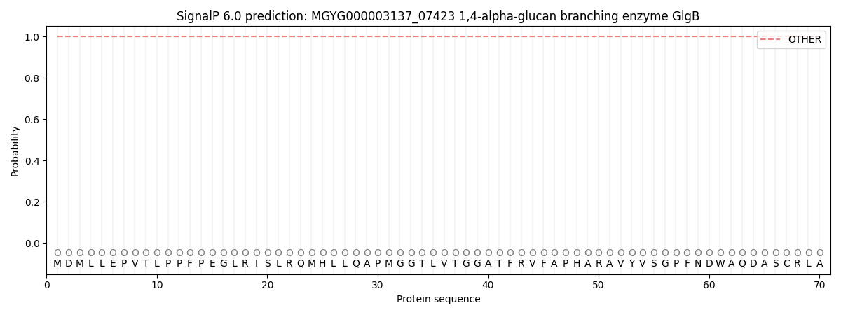
This protein is predicted as OTHER

| Other | SP_Sec_SPI | LIPO_Sec_SPII | TAT_Tat_SPI | TATLIP_Sec_SPII | PILIN_Sec_SPIII |
|---|---|---|---|---|---|
| 1.000048 | 0.000000 | 0.000000 | 0.000000 | 0.000000 | 0.000000 |
