You are browsing environment: HUMAN GUT
CAZyme Information: MGYG000003193_02053
You are here: Home > Sequence: MGYG000003193_02053
Basic Information |
Genomic context |
Full Sequence |
Enzyme annotations |
CAZy signature domains |
CDD domains |
CAZyme hits |
PDB hits |
Swiss-Prot hits |
SignalP and Lipop annotations |
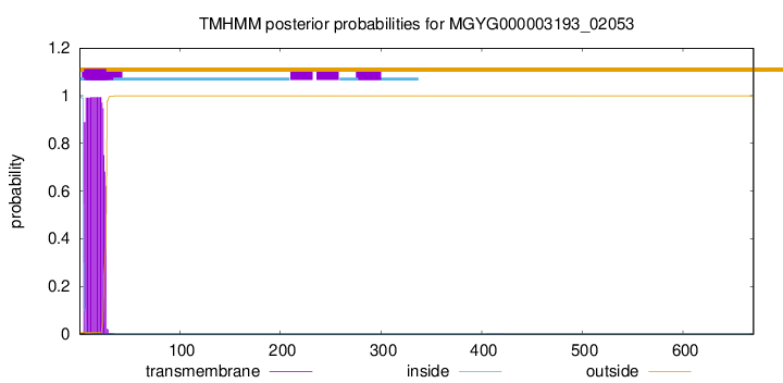
TMHMM annotations
Basic Information help
| Species | CAG-831 sp900544175 | |||||||||||
|---|---|---|---|---|---|---|---|---|---|---|---|---|
| Lineage | Bacteria; Bacteroidota; Bacteroidia; Bacteroidales; UBA932; CAG-831; CAG-831 sp900544175 | |||||||||||
| CAZyme ID | MGYG000003193_02053 | |||||||||||
| CAZy Family | GH20 | |||||||||||
| CAZyme Description | hypothetical protein | |||||||||||
| CAZyme Property |
|
|||||||||||
| Genome Property |
|
|||||||||||
| Gene Location | Start: 4386; End: 6398 Strand: + | |||||||||||
CAZyme Signature Domains help
| Family | Start | End | Evalue | family coverage |
|---|---|---|---|---|
| GH20 | 169 | 490 | 6.4e-69 | 0.9643916913946587 |
CDD Domains download full data without filtering help
| Cdd ID | Domain | E-Value | qStart | qEnd | sStart | sEnd | Domain Description |
|---|---|---|---|---|---|---|---|
| cd06564 | GH20_DspB_LnbB-like | 9.19e-127 | 174 | 490 | 3 | 326 | Glycosyl hydrolase family 20 (GH20) catalytic domain of dispersin B (DspB), lacto-N-biosidase (LnbB) and related proteins. Dispersin B is a soluble beta-N-acetylglucosamidase found in bacteria that hydrolyzes the beta-1,6-linkages of PGA (poly-beta-(1,6)-N-acetylglucosamine), a major component of the extracellular polysaccharide matrix. Lacto-N-biosidase hydrolyzes lacto-N-biose (LNB) type I oligosaccharides at the nonreducing terminus to produce lacto-N-biose as part of the GNB/LNB (galacto-N-biose/lacto-N-biose I) degradation pathway. The lacto-N-biosidase from Bifidobacterium bifidum has this GH20 domain, a carbohydrate binding module 32, and a bacterial immunoglobulin-like domain 2, as well as a YSIRK signal peptide and a G5 membrane anchor at the N and C termini, respectively. The GH20 hexosaminidases are thought to act via a catalytic mechanism in which the catalytic nucleophile is not provided by solvent or the enzyme, but by the substrate itself. |
| cd02742 | GH20_hexosaminidase | 1.79e-42 | 173 | 488 | 1 | 302 | Beta-N-acetylhexosaminidases of glycosyl hydrolase family 20 (GH20) catalyze the removal of beta-1,4-linked N-acetyl-D-hexosamine residues from the non-reducing ends of N-acetyl-beta-D-hexosaminides including N-acetylglucosides and N-acetylgalactosides. These enzymes are broadly distributed in microorganisms, plants and animals, and play roles in various key physiological and pathological processes. These processes include cell structural integrity, energy storage, cellular signaling, fertilization, pathogen defense, viral penetration, the development of carcinomas, inflammatory events and lysosomal storage disorders. The GH20 enzymes include the eukaryotic beta-N-acetylhexosaminidases A and B, the bacterial chitobiases, dispersin B, and lacto-N-biosidase. The GH20 hexosaminidases are thought to act via a catalytic mechanism in which the catalytic nucleophile is not provided by the solvent or the enzyme, but by the substrate itself. |
| pfam00728 | Glyco_hydro_20 | 8.14e-40 | 171 | 490 | 1 | 345 | Glycosyl hydrolase family 20, catalytic domain. This domain has a TIM barrel fold. |
| cd06563 | GH20_chitobiase-like | 5.05e-37 | 171 | 492 | 1 | 346 | The chitobiase of Serratia marcescens is a beta-N-1,4-acetylhexosaminidase with a glycosyl hydrolase family 20 (GH20) domain that hydrolyzes the beta-1,4-glycosidic linkages in oligomers derived from chitin. Chitin is degraded by a two step process: i) a chitinase hydrolyzes the chitin to oligosaccharides and disaccharides such as di-N-acetyl-D-glucosamine and chitobiose, ii) chitobiase then further degrades these oligomers into monomers. This GH20 domain family includes an N-acetylglucosamidase (GlcNAcase A) from Pseudoalteromonas piscicida and an N-acetylhexosaminidase (SpHex) from Streptomyces plicatus. SpHex lacks the C-terminal PKD (polycystic kidney disease I)-like domain found in the chitobiases. The GH20 hexosaminidases are thought to act via a catalytic mechanism in which the catalytic nucleophile is not provided by solvent or the enzyme, but by the substrate itself. |
| COG3525 | Chb | 2.44e-35 | 50 | 489 | 147 | 614 | N-acetyl-beta-hexosaminidase [Carbohydrate transport and metabolism]. |
CAZyme Hits help
| Hit ID | E-Value | Query Start | Query End | Hit Start | Hit End |
|---|---|---|---|---|---|
| QCQ35826.1 | 5.58e-315 | 6 | 665 | 7 | 650 |
| QRM69715.1 | 5.58e-315 | 6 | 665 | 7 | 650 |
| QCQ53716.1 | 5.58e-315 | 6 | 665 | 7 | 650 |
| QCQ49200.1 | 5.58e-315 | 6 | 665 | 7 | 650 |
| QTO27252.1 | 5.58e-315 | 6 | 665 | 7 | 650 |
PDB Hits download full data without filtering help
| Hit ID | E-Value | Query Start | Query End | Hit Start | Hit End | Description |
|---|---|---|---|---|---|---|
| 6JQF_A | 9.12e-82 | 24 | 620 | 82 | 678 | Crystallizationanalysis of a beta-N-acetylhexosaminidase (Am2136) from Akkermansia muciniphila [Akkermansia muciniphila ATCC BAA-835] |
| 3GH4_A | 2.60e-26 | 23 | 490 | 31 | 492 | Crystalstructure of beta-hexosaminidase from Paenibacillus sp. TS12 [Paenibacillus sp.],3GH5_A Crystal structure of beta-hexosaminidase from Paenibacillus sp. TS12 in complex with GlcNAc [Paenibacillus sp.],3GH7_A Crystal structure of beta-hexosaminidase from Paenibacillus sp. TS12 in complex with GalNAc [Paenibacillus sp.],3SUR_A Crystal structure of beta-hexosaminidase from Paenibacillus sp. TS12 in complex with NAG-thiazoline. [Paenibacillus sp. TS12],3SUS_A Crystal structure of beta-hexosaminidase from Paenibacillus sp. TS12 in complex with Gal-NAG-thiazoline [Paenibacillus sp. TS12],3SUT_A Crystal structure of beta-hexosaminidase from Paenibacillus sp. TS12 in complex with PUGNAc [Paenibacillus sp. TS12],3SUU_A Crystal structure of beta-hexosaminidase from Paenibacillus sp. TS12 in complex with Gal-PUGNAc [Paenibacillus sp. TS12],3SUV_A Crystal structure of beta-hexosaminidase from Paenibacillus sp. TS12 in complex with NHAc-DNJ [Paenibacillus sp. TS12],3SUW_A Crystal structure of beta-hexosaminidase from Paenibacillus sp. TS12 in complex with NHAc-CAS [Paenibacillus sp. TS12] |
| 6YHH_A | 1.70e-25 | 114 | 490 | 92 | 478 | X-rayStructure of Flavobacterium johnsoniae chitobiase (FjGH20) [Flavobacterium johnsoniae UW101],6YHH_B X-ray Structure of Flavobacterium johnsoniae chitobiase (FjGH20) [Flavobacterium johnsoniae UW101] |
| 7BWG_A | 6.58e-25 | 23 | 530 | 10 | 518 | AGlycoside Hydrolase Family 20 beta-N-Acetylglucosaminidase [Microbacterium sp. HJ5],7BWG_B A Glycoside Hydrolase Family 20 beta-N-Acetylglucosaminidase [Microbacterium sp. HJ5] |
| 3RCN_A | 1.66e-23 | 113 | 490 | 77 | 485 | CrystalStructure of Beta-N-Acetylhexosaminidase from Arthrobacter aurescens [Paenarthrobacter aurescens TC1] |
Swiss-Prot Hits download full data without filtering help
| Hit ID | E-Value | Query Start | Query End | Hit Start | Hit End | Description |
|---|---|---|---|---|---|---|
| B2UPR7 | 5.25e-85 | 24 | 620 | 104 | 700 | Beta-hexosaminidase Amuc_2136 OS=Akkermansia muciniphila (strain ATCC BAA-835 / DSM 22959 / JCM 33894 / BCRC 81048 / CCUG 64013 / CIP 107961 / Muc) OX=349741 GN=Amuc_2136 PE=1 SV=1 |
| Q54SC9 | 6.74e-23 | 106 | 492 | 101 | 480 | Beta-hexosaminidase subunit A2 OS=Dictyostelium discoideum OX=44689 GN=hexa2 PE=3 SV=1 |
| B2UP57 | 1.55e-19 | 87 | 332 | 34 | 279 | Beta-hexosaminidase Amuc_2018 OS=Akkermansia muciniphila (strain ATCC BAA-835 / DSM 22959 / JCM 33894 / BCRC 81048 / CCUG 64013 / CIP 107961 / Muc) OX=349741 GN=Amuc_2018 PE=1 SV=1 |
| Q7WUL4 | 1.55e-18 | 33 | 492 | 6 | 452 | Beta-N-acetylhexosaminidase OS=Cellulomonas fimi OX=1708 GN=hex20 PE=1 SV=1 |
| Q5RC84 | 2.40e-18 | 112 | 494 | 112 | 491 | Beta-hexosaminidase subunit alpha OS=Pongo abelii OX=9601 GN=HEXA PE=3 SV=1 |
SignalP and Lipop Annotations help
This protein is predicted as SP
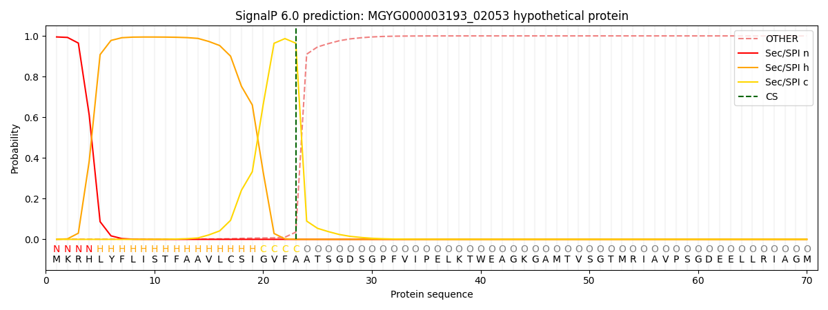
| Other | SP_Sec_SPI | LIPO_Sec_SPII | TAT_Tat_SPI | TATLIP_Sec_SPII | PILIN_Sec_SPIII |
|---|---|---|---|---|---|
| 0.001989 | 0.992582 | 0.004710 | 0.000286 | 0.000220 | 0.000200 |