You are browsing environment: HUMAN GUT
CAZyme Information: MGYG000003293_00015
You are here: Home > Sequence: MGYG000003293_00015
Basic Information |
Genomic context |
Full Sequence |
Enzyme annotations |
CAZy signature domains |
CDD domains |
CAZyme hits |
PDB hits |
Swiss-Prot hits |
SignalP and Lipop annotations |
TMHMM annotations
Basic Information help
| Species | CAG-110 sp900544405 | |||||||||||
|---|---|---|---|---|---|---|---|---|---|---|---|---|
| Lineage | Bacteria; Firmicutes_A; Clostridia; Oscillospirales; Oscillospiraceae; CAG-110; CAG-110 sp900544405 | |||||||||||
| CAZyme ID | MGYG000003293_00015 | |||||||||||
| CAZy Family | GT28 | |||||||||||
| CAZyme Description | Processive diacylglycerol beta-glucosyltransferase | |||||||||||
| CAZyme Property |
|
|||||||||||
| Genome Property |
|
|||||||||||
| Gene Location | Start: 15434; End: 17173 Strand: - | |||||||||||
CAZyme Signature Domains help
| Family | Start | End | Evalue | family coverage |
|---|---|---|---|---|
| GT28 | 205 | 349 | 2.3e-28 | 0.9490445859872612 |
CDD Domains download full data without filtering help
| Cdd ID | Domain | E-Value | qStart | qEnd | sStart | sEnd | Domain Description |
|---|---|---|---|---|---|---|---|
| cd17507 | GT28_Beta-DGS-like | 1.14e-62 | 3 | 360 | 1 | 356 | beta-diglucosyldiacylglycerol synthase and similar proteins. beta-diglucosyldiacylglycerol synthase (processive diacylglycerol beta-glucosyltransferase EC 2.4.1.315) is involved in the biosynthesis of both the bilayer- and non-bilayer-forming membrane glucolipids. This family of glycosyltransferases also contains plant major galactolipid synthase (chloroplastic monogalactosyldiacylglycerol synthase 1 EC 2.4.1.46). Glycosyltransferases catalyze the transfer of sugar moieties from activated donor molecules to specific acceptor molecules, forming glycosidic bonds. The acceptor molecule can be a lipid, a protein, a heterocyclic compound, or another carbohydrate residue. The structures of the formed glycoconjugates are extremely diverse, reflecting a wide range of biological functions. The members of this family share a common GTB topology, one of the two protein topologies observed for nucleotide-sugar-dependent glycosyltransferases. GTB proteins have distinct N- and C- terminal domains each containing a typical Rossmann fold. The two domains have high structural homology despite minimal sequence homology. The large cleft that separates the two domains includes the catalytic center and permits a high degree of flexibility. |
| cd01041 | Rubrerythrin | 1.49e-47 | 397 | 531 | 1 | 129 | Rubrerythrin, ferritin-like diiron-binding domain. Rubrerythrin domain is a nonheme iron binding domain found in many air-sensitive bacteria and archaea and member of a broad superfamily of ferritin-like diiron-carboxylate proteins. The homodimeric rubrerythrin protein contains a binuclear metal center located within a four helix bundle. Many, but not all, rubrerythrin proteins have a second domain with a rubredoxin-like hexacoordinated iron center. Rubrerythrin is thought to reduce hydrogen peroxide as part of an oxidative stress protection system but its function is still poorly understood. |
| COG1592 | YotD | 5.73e-41 | 396 | 567 | 3 | 156 | Rubrerythrin [Energy production and conversion]. |
| PRK13609 | PRK13609 | 3.96e-39 | 2 | 359 | 6 | 362 | diacylglycerol glucosyltransferase; Provisional |
| PLN02605 | PLN02605 | 8.62e-34 | 3 | 359 | 1 | 372 | monogalactosyldiacylglycerol synthase |
CAZyme Hits help
| Hit ID | E-Value | Query Start | Query End | Hit Start | Hit End |
|---|---|---|---|---|---|
| BCK81078.1 | 9.38e-86 | 1 | 343 | 1 | 348 |
| BCK81488.1 | 2.09e-82 | 1 | 358 | 1 | 357 |
| QUO34454.1 | 1.48e-78 | 1 | 358 | 1 | 357 |
| BCK79763.1 | 3.88e-78 | 1 | 362 | 1 | 362 |
| QNL44772.1 | 1.67e-74 | 1 | 364 | 1 | 365 |
PDB Hits download full data without filtering help
| Hit ID | E-Value | Query Start | Query End | Hit Start | Hit End | Description |
|---|---|---|---|---|---|---|
| 1RYT_A | 3.00e-28 | 397 | 566 | 7 | 176 | Rubrerythrin[Desulfovibrio vulgaris str. Hildenborough] |
| 1B71_A | 3.08e-28 | 397 | 566 | 8 | 177 | Rubrerythrin[Desulfovibrio vulgaris str. Hildenborough],1DVB_A Rubrerythrin [Desulfovibrio vulgaris str. Hildenborough],1JYB_A Crystal structure of Rubrerythrin [Desulfovibrio vulgaris],1LKM_A Crystal structure of Desulfovibrio vulgaris rubrerythrin all-iron(III) form [Desulfovibrio vulgaris],1LKO_A Crystal structure of Desulfovibrio vulgaris rubrerythrin all-iron(II) form [Desulfovibrio vulgaris],1LKP_A Crystal structure of Desulfovibrio vulgaris rubrerythrin all-iron(II) form, azide adduct [Desulfovibrio vulgaris],1QYB_A X-ray crystal structure of Desulfovibrio vulgaris rubrerythrin with zinc substituted into the [Fe(SCys)4] site and alternative diiron site structures [Desulfovibrio vulgaris str. Hildenborough],1S2Z_A X-ray crystal structure of Desulfovibrio vulgaris Rubrerythrin with displacement of iron by zinc at the diiron Site [Desulfovibrio vulgaris],1S30_A X-ray crystal structure of Desulfovibrio vulgaris Rubrerythrin with displacement of iron by zinc at the diiron Site [Desulfovibrio vulgaris] |
| 4WYI_A | 2.75e-27 | 2 | 345 | 7 | 363 | Thecrystal structure of Arabidopsis thaliana galactolipid synthase, MGD1 (apo-form) [Arabidopsis thaliana],4X1T_A The crystal structure of Arabidopsis thaliana galactolipid synthase MGD1 in complex with UDP [Arabidopsis thaliana] |
| 4DI0_A | 1.50e-17 | 397 | 524 | 12 | 131 | Thestructure of Rubrerythrin from Burkholderia pseudomallei [Burkholderia pseudomallei 1710b],4DI0_B The structure of Rubrerythrin from Burkholderia pseudomallei [Burkholderia pseudomallei 1710b] |
| 3MPS_A | 6.22e-16 | 402 | 567 | 11 | 160 | PeroxideBound Oxidized Rubrerythrin from Pyrococcus furiosus [Pyrococcus furiosus],3MPS_B Peroxide Bound Oxidized Rubrerythrin from Pyrococcus furiosus [Pyrococcus furiosus],3MPS_D Peroxide Bound Oxidized Rubrerythrin from Pyrococcus furiosus [Pyrococcus furiosus],3MPS_F Peroxide Bound Oxidized Rubrerythrin from Pyrococcus furiosus [Pyrococcus furiosus],3MPS_G Peroxide Bound Oxidized Rubrerythrin from Pyrococcus furiosus [Pyrococcus furiosus],3MPS_H Peroxide Bound Oxidized Rubrerythrin from Pyrococcus furiosus [Pyrococcus furiosus],3MPS_I Peroxide Bound Oxidized Rubrerythrin from Pyrococcus furiosus [Pyrococcus furiosus],3MPS_K Peroxide Bound Oxidized Rubrerythrin from Pyrococcus furiosus [Pyrococcus furiosus],3PWF_A High resolution structure of the fully reduced form of rubrerythrin from P. furiosus [Pyrococcus furiosus],3PWF_B High resolution structure of the fully reduced form of rubrerythrin from P. furiosus [Pyrococcus furiosus],3PZA_A Fully Reduced (All-ferrous) Pyrococcus rubrerythrin after a 10 second exposure to peroxide. [Pyrococcus furiosus],3PZA_B Fully Reduced (All-ferrous) Pyrococcus rubrerythrin after a 10 second exposure to peroxide. [Pyrococcus furiosus],3QVD_A Exposure of rubrerythrin from Pyrococcus furiosus to peroxide, fifteen second time point. [Pyrococcus furiosus],3QVD_B Exposure of rubrerythrin from Pyrococcus furiosus to peroxide, fifteen second time point. [Pyrococcus furiosus],3QVD_C Exposure of rubrerythrin from Pyrococcus furiosus to peroxide, fifteen second time point. [Pyrococcus furiosus],3QVD_D Exposure of rubrerythrin from Pyrococcus furiosus to peroxide, fifteen second time point. [Pyrococcus furiosus],3QVD_E Exposure of rubrerythrin from Pyrococcus furiosus to peroxide, fifteen second time point. [Pyrococcus furiosus],3QVD_F Exposure of rubrerythrin from Pyrococcus furiosus to peroxide, fifteen second time point. [Pyrococcus furiosus],3QVD_G Exposure of rubrerythrin from Pyrococcus furiosus to peroxide, fifteen second time point. [Pyrococcus furiosus],3QVD_H Exposure of rubrerythrin from Pyrococcus furiosus to peroxide, fifteen second time point. [Pyrococcus furiosus] |
Swiss-Prot Hits download full data without filtering help
| Hit ID | E-Value | Query Start | Query End | Hit Start | Hit End | Description |
|---|---|---|---|---|---|---|
| A8FED1 | 6.53e-35 | 2 | 358 | 6 | 361 | Processive diacylglycerol beta-glucosyltransferase OS=Bacillus pumilus (strain SAFR-032) OX=315750 GN=ugtP PE=3 SV=1 |
| P54166 | 4.24e-34 | 2 | 358 | 6 | 361 | Processive diacylglycerol beta-glucosyltransferase OS=Bacillus subtilis (strain 168) OX=224308 GN=ugtP PE=1 SV=1 |
| P51591 | 7.97e-33 | 397 | 566 | 8 | 181 | Rubrerythrin OS=Clostridium perfringens (strain 13 / Type A) OX=195102 GN=rbr PE=3 SV=2 |
| Q9AGG3 | 1.73e-32 | 397 | 575 | 17 | 195 | Rubrerythrin OS=Porphyromonas gingivalis (strain ATCC BAA-308 / W83) OX=242619 GN=rbr PE=2 SV=1 |
| Q65IA4 | 3.45e-32 | 3 | 358 | 7 | 361 | Processive diacylglycerol beta-glucosyltransferase OS=Bacillus licheniformis (strain ATCC 14580 / DSM 13 / JCM 2505 / CCUG 7422 / NBRC 12200 / NCIMB 9375 / NCTC 10341 / NRRL NRS-1264 / Gibson 46) OX=279010 GN=ugtP PE=3 SV=1 |
SignalP and Lipop Annotations help
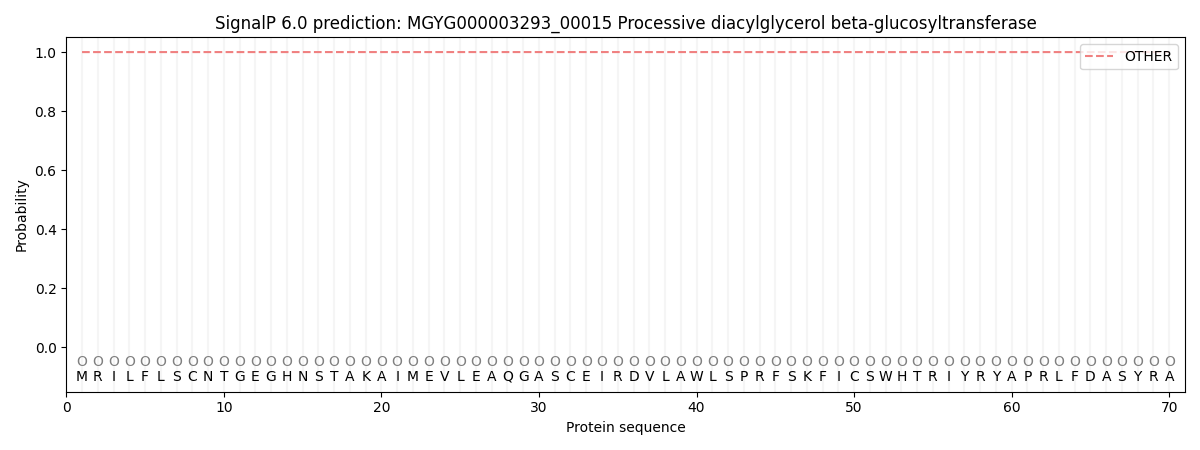
This protein is predicted as OTHER

| Other | SP_Sec_SPI | LIPO_Sec_SPII | TAT_Tat_SPI | TATLIP_Sec_SPII | PILIN_Sec_SPIII |
|---|---|---|---|---|---|
| 1.000060 | 0.000000 | 0.000000 | 0.000000 | 0.000000 | 0.000000 |
