You are browsing environment: HUMAN GUT
CAZyme Information: MGYG000003468_00145
You are here: Home > Sequence: MGYG000003468_00145
Basic Information |
Genomic context |
Full Sequence |
Enzyme annotations |
CAZy signature domains |
CDD domains |
CAZyme hits |
PDB hits |
Swiss-Prot hits |
SignalP and Lipop annotations |
TMHMM annotations
Basic Information help
| Species | UMGS1883 sp900768005 | |||||||||||
|---|---|---|---|---|---|---|---|---|---|---|---|---|
| Lineage | Bacteria; Firmicutes_A; Clostridia; UMGS1883; UMGS1883; UMGS1883; UMGS1883 sp900768005 | |||||||||||
| CAZyme ID | MGYG000003468_00145 | |||||||||||
| CAZy Family | CBM50 | |||||||||||
| CAZyme Description | hypothetical protein | |||||||||||
| CAZyme Property |
|
|||||||||||
| Genome Property |
|
|||||||||||
| Gene Location | Start: 1239; End: 2084 Strand: + | |||||||||||
CDD Domains download full data without filtering help
| Cdd ID | Domain | E-Value | qStart | qEnd | sStart | sEnd | Domain Description |
|---|---|---|---|---|---|---|---|
| cd16913 | YkuD_like | 8.62e-42 | 175 | 279 | 2 | 120 | L,D-transpeptidases/carboxypeptidases similar to Bacillus YkuD. Members of the YkuD-like family of proteins are found in a range of bacteria. The best studied member Bacillus YkuD has been shown to act as an L,D-transpeptidase that gives rise to an alternative pathway for peptidoglycan cross-linking. Another member Helicobacter pylori Csd6 functions as an L,D-carboxypeptidase and regulates helical cell shape and motility. The conserved region contains a conserved histidine and cysteine, with the cysteine thought to be an active site residue. |
| COG1376 | ErfK | 2.68e-24 | 147 | 279 | 78 | 228 | Lipoprotein-anchoring transpeptidase ErfK/SrfK [Cell wall/membrane/envelope biogenesis]. |
| pfam03734 | YkuD | 9.82e-24 | 172 | 279 | 1 | 89 | L,D-transpeptidase catalytic domain. This family of proteins are found in a range of bacteria. It has been shown that this domain can act as an L,D-transpeptidase that gives rise to an alternative pathway for peptidoglycan cross-linking. This gives bacteria resistance to beta-lactam antibiotics that inhibit PBPs which usually carry out the cross-linking reaction. The conserved region contains a conserved histidine and cysteine, with the cysteine thought to be an active site residue. Several members of this family contain peptidoglycan binding domains. The molecular structure of YkuD protein shows this domain has a novel tertiary fold consisting of a beta-sandwich with two mixed sheets, one containing five strands and the other, six strands. The two beta-sheets form a cradle capped by an alpha-helix. This family was formerly called the ErfK/YbiS/YcfS/YnhG family, but is now named after the first protein of known structure. |
| smart00257 | LysM | 3.31e-13 | 6 | 49 | 1 | 44 | Lysin motif. |
| smart00257 | LysM | 4.58e-13 | 66 | 108 | 2 | 44 | Lysin motif. |
CAZyme Hits help
| Hit ID | E-Value | Query Start | Query End | Hit Start | Hit End |
|---|---|---|---|---|---|
| ALX07568.1 | 7.45e-109 | 7 | 279 | 16 | 288 |
| ANV75308.1 | 7.45e-109 | 7 | 279 | 16 | 288 |
| ADU73640.1 | 7.45e-109 | 7 | 279 | 16 | 288 |
| ABN54202.1 | 1.06e-108 | 7 | 279 | 16 | 288 |
| AOT72145.1 | 2.90e-96 | 7 | 279 | 17 | 288 |
PDB Hits download full data without filtering help
| Hit ID | E-Value | Query Start | Query End | Hit Start | Hit End | Description |
|---|---|---|---|---|---|---|
| 4A1I_A | 1.82e-38 | 124 | 279 | 5 | 163 | ykudfrom B.subtilis [Bacillus subtilis],4A1I_B ykud from B.subtilis [Bacillus subtilis],4A1I_C ykud from B.subtilis [Bacillus subtilis],4A1I_D ykud from B.subtilis [Bacillus subtilis],4A1I_E ykud from B.subtilis [Bacillus subtilis],4A1I_F ykud from B.subtilis [Bacillus subtilis],4A1I_G ykud from B.subtilis [Bacillus subtilis],4A1I_H ykud from B.subtilis [Bacillus subtilis],4A1J_A Ykud L,D-transpeptidase from B.subtilis [Bacillus subtilis],4A1J_B Ykud L,D-transpeptidase from B.subtilis [Bacillus subtilis] |
| 2MTZ_A | 6.83e-38 | 124 | 279 | 7 | 165 | Haddockmodel of Bacillus subtilis L,D-transpeptidase in complex with a peptidoglycan hexamuropeptide [Bacillus subtilis],3ZQD_A B. subtilis L,D-transpeptidase [Bacillus subtilis subsp. subtilis str. 168],4A52_A NMR structure of the imipenem-acylated L,D-transpeptidase from Bacillus subtilis [Bacillus subtilis subsp. subtilis str. 168] |
| 1Y7M_A | 1.96e-37 | 124 | 279 | 4 | 162 | ChainA, Crystal Structure of the B. subtilis YkuD protein at 2 A resolution [Bacillus subtilis subsp. subtilis str. 168],1Y7M_B Chain B, Crystal Structure of the B. subtilis YkuD protein at 2 A resolution [Bacillus subtilis subsp. subtilis str. 168] |
| 4A1K_A | 5.64e-37 | 124 | 279 | 5 | 163 | YkudL,D-transpeptidase [Bacillus subtilis] |
| 4B8V_A | 3.12e-16 | 7 | 167 | 44 | 217 | ChainA, Extracellular Protein 6 [Fulvia fulva],4B9H_A Chain A, Extracellular Protein 6 [Fulvia fulva] |
Swiss-Prot Hits download full data without filtering help
| Hit ID | E-Value | Query Start | Query End | Hit Start | Hit End | Description |
|---|---|---|---|---|---|---|
| Q5WC42 | 2.32e-45 | 123 | 281 | 3 | 165 | Putative L,D-transpeptidase YkuD OS=Alkalihalobacillus clausii (strain KSM-K16) OX=66692 GN=ABC3535 PE=3 SV=1 |
| O34816 | 2.72e-37 | 124 | 279 | 4 | 162 | Putative L,D-transpeptidase YkuD OS=Bacillus subtilis (strain 168) OX=224308 GN=ykuD PE=1 SV=1 |
| Q65K99 | 7.83e-37 | 124 | 281 | 4 | 165 | Putative L,D-transpeptidase YkuD OS=Bacillus licheniformis (strain ATCC 14580 / DSM 13 / JCM 2505 / CCUG 7422 / NBRC 12200 / NCIMB 9375 / NCTC 10341 / NRRL NRS-1264 / Gibson 46) OX=279010 GN=BLi01617 PE=3 SV=1 |
| P54539 | 1.23e-18 | 170 | 279 | 24 | 150 | Putative L,D-transpeptidase YqjB OS=Bacillus subtilis (strain 168) OX=224308 GN=yqjB PE=3 SV=1 |
| P76193 | 6.50e-10 | 124 | 280 | 42 | 233 | Probable L,D-transpeptidase YnhG OS=Escherichia coli (strain K12) OX=83333 GN=ynhG PE=1 SV=1 |
SignalP and Lipop Annotations help
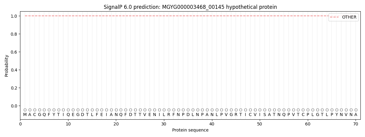
This protein is predicted as OTHER
| Other | SP_Sec_SPI | LIPO_Sec_SPII | TAT_Tat_SPI | TATLIP_Sec_SPII | PILIN_Sec_SPIII |
|---|---|---|---|---|---|
| 1.000060 | 0.000019 | 0.000001 | 0.000000 | 0.000000 | 0.000000 |