You are browsing environment: HUMAN GUT
CAZyme Information: MGYG000003595_00189
You are here: Home > Sequence: MGYG000003595_00189
Basic Information |
Genomic context |
Full Sequence |
Enzyme annotations |
CAZy signature domains |
CDD domains |
CAZyme hits |
PDB hits |
Swiss-Prot hits |
SignalP and Lipop annotations |
TMHMM annotations
Basic Information help
| Species | HGM11525 sp900770405 | |||||||||||
|---|---|---|---|---|---|---|---|---|---|---|---|---|
| Lineage | Bacteria; Firmicutes_A; Clostridia; Monoglobales_A; UBA1381; HGM11525; HGM11525 sp900770405 | |||||||||||
| CAZyme ID | MGYG000003595_00189 | |||||||||||
| CAZy Family | GH151 | |||||||||||
| CAZyme Description | hypothetical protein | |||||||||||
| CAZyme Property |
|
|||||||||||
| Genome Property |
|
|||||||||||
| Gene Location | Start: 13656; End: 15620 Strand: - | |||||||||||
CAZyme Signature Domains help
| Family | Start | End | Evalue | family coverage |
|---|---|---|---|---|
| GH151 | 25 | 155 | 9.4e-51 | 0.9847328244274809 |
CDD Domains download full data without filtering help
| Cdd ID | Domain | E-Value | qStart | qEnd | sStart | sEnd | Domain Description |
|---|---|---|---|---|---|---|---|
| pfam14871 | GHL6 | 1.63e-27 | 24 | 155 | 1 | 135 | Hypothetical glycosyl hydrolase 6. GHL6 is a family of hypothetical glycoside hydrolases. |
| cd03143 | A4_beta-galactosidase_middle_domain | 3.75e-07 | 372 | 429 | 33 | 90 | A4 beta-galactosidase middle domain: a type 1 glutamine amidotransferase (GATase1)-like domain. A4 beta-galactosidase middle domain: a type 1 glutamine amidotransferase (GATase1)-like domain. This group includes proteins similar to beta-galactosidase from Thermus thermophilus. Beta-Galactosidase hydrolyzes the beta-1,4-D-galactosidic linkage of lactose, as well as those of related chromogens, o-nitrophenyl-beta-D-galactopyranoside (ONP-Gal) and 5-bromo-4-chloro-3-indolyl-beta-D-galactoside (X-gal). This A4 beta-galactosidase middle domain lacks the catalytic triad of typical GATase1 domains. The reactive Cys residue found in the sharp turn between a beta strand and an alpha helix termed the nucleophile elbow in typical GATase1 domains is not conserved in this group. |
| pfam08532 | Glyco_hydro_42M | 0.002 | 382 | 429 | 47 | 94 | Beta-galactosidase trimerisation domain. This is non catalytic domain B of beta-galactosidase enzymes belong to the glycosyl hydrolase 42 family. This domain is related to glutamine amidotransferase enzymes, but the catalytic residues are replaced by non functional amino acids. This domain is involved in trimerisation. |
| COG1874 | GanA | 0.004 | 381 | 464 | 446 | 520 | Beta-galactosidase GanA [Carbohydrate transport and metabolism]. |
| cd05269 | TMR_SDR_a | 0.009 | 73 | 187 | 85 | 179 | triphenylmethane reductase (TMR)-like proteins, NMRa-like, atypical (a) SDRs. TMR is an atypical NADP-binding protein of the SDR family. It lacks the active site residues of the SDRs but has a glycine rich NAD(P)-binding motif that matches the extended SDRs. Proteins in this subgroup however, are more similar in length to the classical SDRs. TMR was identified as a reducer of triphenylmethane dyes, important environmental pollutants. This subgroup also includes Escherichia coli NADPH-dependent quinine oxidoreductase (QOR2), which catalyzes two-electron reduction of quinone; but is unlikely to play a major role in protecting against quinone cytotoxicity. Atypical SDRs are distinct from classical SDRs. Atypical SDRs include biliverdin IX beta reductase (BVR-B,aka flavin reductase), NMRa (a negative transcriptional regulator of various fungi), progesterone 5-beta-reductase like proteins, phenylcoumaran benzylic ether and pinoresinol-lariciresinol reductases, phenylpropene synthases, eugenol synthase, triphenylmethane reductase, isoflavone reductases, and others. SDRs are a functionally diverse family of oxidoreductases that have a single domain with a structurally conserved Rossmann fold, an NAD(P)(H)-binding region, and a structurally diverse C-terminal region. Sequence identity between different SDR enzymes is typically in the 15-30% range; they catalyze a wide range of activities including the metabolism of steroids, cofactors, carbohydrates, lipids, aromatic compounds, and amino acids, and act in redox sensing. Classical SDRs have an TGXXX[AG]XG cofactor binding motif and a YXXXK active site motif, with the Tyr residue of the active site motif serving as a critical catalytic residue (Tyr-151, human 15-hydroxyprostaglandin dehydrogenase numbering). In addition to the Tyr and Lys, there is often an upstream Ser and/or an Asn, contributing to the active site; while substrate binding is in the C-terminal region, which determines specificity. The standard reaction mechanism is a 4-pro-S hydride transfer and proton relay involving the conserved Tyr and Lys, a water molecule stabilized by Asn, and nicotinamide. In addition to the Rossmann fold core region typical of all SDRs, extended SDRs have a less conserved C-terminal extension of approximately 100 amino acids, and typically have a TGXXGXXG cofactor binding motif. Complex (multidomain) SDRs such as ketoreductase domains of fatty acid synthase have a GGXGXXG NAD(P)-binding motif and an altered active site motif (YXXXN). Fungal type ketoacyl reductases have a TGXXXGX(1-2)G NAD(P)-binding motif. |
CAZyme Hits help
| Hit ID | E-Value | Query Start | Query End | Hit Start | Hit End |
|---|---|---|---|---|---|
| BCJ93865.1 | 2.15e-271 | 1 | 654 | 1 | 659 |
| BCJ98360.1 | 5.70e-271 | 1 | 654 | 1 | 657 |
| QNK60374.1 | 1.65e-265 | 1 | 653 | 1 | 654 |
| QHT63046.1 | 7.25e-261 | 1 | 654 | 1 | 663 |
| AFH63272.1 | 1.22e-260 | 1 | 654 | 1 | 658 |
PDB Hits download full data without filtering help
| Hit ID | E-Value | Query Start | Query End | Hit Start | Hit End | Description |
|---|---|---|---|---|---|---|
| 6TVK_AAA | 9.69e-239 | 1 | 654 | 30 | 686 | ChainAAA, Alpha-L-fucosidase [Paenibacillus thiaminolyticus] |
Swiss-Prot Hits help
SignalP and Lipop Annotations help
This protein is predicted as OTHER
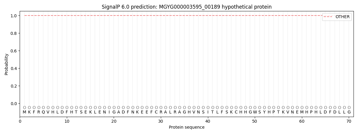
| Other | SP_Sec_SPI | LIPO_Sec_SPII | TAT_Tat_SPI | TATLIP_Sec_SPII | PILIN_Sec_SPIII |
|---|---|---|---|---|---|
| 1.000075 | 0.000000 | 0.000000 | 0.000000 | 0.000000 | 0.000000 |