You are browsing environment: HUMAN GUT
CAZyme Information: MGYG000003761_01246
You are here: Home > Sequence: MGYG000003761_01246
Basic Information |
Genomic context |
Full Sequence |
Enzyme annotations |
CAZy signature domains |
CDD domains |
CAZyme hits |
PDB hits |
Swiss-Prot hits |
SignalP and Lipop annotations |
TMHMM annotations
Basic Information help
| Species | Stenotrophomonas maltophilia_S | |||||||||||
|---|---|---|---|---|---|---|---|---|---|---|---|---|
| Lineage | Bacteria; Proteobacteria; Gammaproteobacteria; Xanthomonadales; Xanthomonadaceae; Stenotrophomonas; Stenotrophomonas maltophilia_S | |||||||||||
| CAZyme ID | MGYG000003761_01246 | |||||||||||
| CAZy Family | GT4 | |||||||||||
| CAZyme Description | D-inositol-3-phosphate glycosyltransferase | |||||||||||
| CAZyme Property |
|
|||||||||||
| Genome Property |
|
|||||||||||
| Gene Location | Start: 9593; End: 12958 Strand: + | |||||||||||
CAZyme Signature Domains help
| Family | Start | End | Evalue | family coverage |
|---|---|---|---|---|
| GT4 | 653 | 804 | 5.7e-23 | 0.9375 |
| GT2 | 839 | 953 | 2.6e-21 | 0.6647058823529411 |
CDD Domains download full data without filtering help
| Cdd ID | Domain | E-Value | qStart | qEnd | sStart | sEnd | Domain Description |
|---|---|---|---|---|---|---|---|
| cd11579 | Glyco_tran_WbsX | 0.0 | 123 | 463 | 4 | 345 | Glycosyl hydrolase family 99-like domain of WbsX-like glycosyltransferases. Members of this domain family are found in proteins within O-antigen biosynthesis clusters in Gram negative bacteria, where they may function as glycosyl hydrolases and typically co-occur with glycosyltransferase domains. They bear resemblance to GH71 and the GH99 family of alpha-1,2-mannosidases and may share a similar cataltyic site and mechanism. The O-antigens are essential lipopolysaccharides in gram-negative bacteria's outer membrane and have been linked to pathogenicity. |
| pfam14307 | Glyco_tran_WbsX | 3.79e-179 | 123 | 460 | 2 | 312 | Glycosyltransferase WbsX. Members of this family are found in within O-antigen biosynthesis clusters in Gram negative bacteria, where they are predicted to function as glycosyltransferases. |
| COG3754 | RgpF | 4.08e-32 | 118 | 467 | 91 | 407 | Lipopolysaccharide biosynthesis protein [Cell wall/membrane/envelope biogenesis]. |
| cd11573 | GH99_GH71_like | 4.01e-31 | 169 | 454 | 2 | 259 | Glycoside hydrolase families 71, 99, and related domains. This superfamily of glycoside hydrolases contains families GH71 and GH99 (following the CAZY nomenclature), as well as other members with undefined function and specificity. |
| cd03801 | GT4_PimA-like | 1.29e-30 | 471 | 830 | 1 | 364 | phosphatidyl-myo-inositol mannosyltransferase. This family is most closely related to the GT4 family of glycosyltransferases and named after PimA in Propionibacterium freudenreichii, which is involved in the biosynthesis of phosphatidyl-myo-inositol mannosides (PIM) which are early precursors in the biosynthesis of lipomannans (LM) and lipoarabinomannans (LAM), and catalyzes the addition of a mannosyl residue from GDP-D-mannose (GDP-Man) to the position 2 of the carrier lipid phosphatidyl-myo-inositol (PI) to generate a phosphatidyl-myo-inositol bearing an alpha-1,2-linked mannose residue (PIM1). Glycosyltransferases catalyze the transfer of sugar moieties from activated donor molecules to specific acceptor molecules, forming glycosidic bonds. The acceptor molecule can be a lipid, a protein, a heterocyclic compound, or another carbohydrate residue. This group of glycosyltransferases is most closely related to the previously defined glycosyltransferase family 1 (GT1). The members of this family may transfer UDP, ADP, GDP, or CMP linked sugars. The diverse enzymatic activities among members of this family reflect a wide range of biological functions. The protein structure available for this family has the GTB topology, one of the two protein topologies observed for nucleotide-sugar-dependent glycosyltransferases. GTB proteins have distinct N- and C- terminal domains each containing a typical Rossmann fold. The two domains have high structural homology despite minimal sequence homology. The large cleft that separates the two domains includes the catalytic center and permits a high degree of flexibility. The members of this family are found mainly in certain bacteria and archaea. |
CAZyme Hits help
| Hit ID | E-Value | Query Start | Query End | Hit Start | Hit End |
|---|---|---|---|---|---|
| QNG85928.1 | 0.0 | 1 | 1121 | 1 | 1122 |
| QNG92428.1 | 0.0 | 1 | 1121 | 1 | 1122 |
| CCH11119.1 | 0.0 | 1 | 1121 | 1 | 1122 |
| AWB76961.1 | 0.0 | 1 | 1121 | 1 | 1122 |
| ATF87117.1 | 0.0 | 1 | 1113 | 545 | 1641 |
PDB Hits download full data without filtering help
| Hit ID | E-Value | Query Start | Query End | Hit Start | Hit End | Description |
|---|---|---|---|---|---|---|
| 3OKA_A | 1.04e-09 | 637 | 838 | 183 | 375 | Crystalstructure of Corynebacterium glutamicum PimB' in complex with GDP-Man (triclinic crystal form) [Corynebacterium glutamicum],3OKA_B Crystal structure of Corynebacterium glutamicum PimB' in complex with GDP-Man (triclinic crystal form) [Corynebacterium glutamicum] |
| 3OKC_A | 1.10e-09 | 637 | 838 | 183 | 375 | Crystalstructure of Corynebacterium glutamicum PimB' bound to GDP (orthorhombic crystal form) [Corynebacterium glutamicum],3OKP_A Crystal structure of Corynebacterium glutamicum PimB' bound to GDP-Man (orthorhombic crystal form) [Corynebacterium glutamicum] |
| 6YV7_B | 1.07e-06 | 836 | 930 | 42 | 138 | MannosyltransferasePcManGT from Pyrobaculum calidifontis [Pyrobaculum calidifontis JCM 11548],6YV8_B Mannosyltransferase PcManGT from Pyrobaculum calidifontis in complex with GDP and Mn2+ [Pyrobaculum calidifontis JCM 11548],6YV9_A Mannosyltransferase PcManGT from Pyrobaculum calidifontis in complex with GDP-Man and Mn2+ [Pyrobaculum calidifontis JCM 11548] |
| 6YV7_A | 1.08e-06 | 836 | 930 | 43 | 139 | MannosyltransferasePcManGT from Pyrobaculum calidifontis [Pyrobaculum calidifontis JCM 11548],6YV8_A Mannosyltransferase PcManGT from Pyrobaculum calidifontis in complex with GDP and Mn2+ [Pyrobaculum calidifontis JCM 11548],6YV9_B Mannosyltransferase PcManGT from Pyrobaculum calidifontis in complex with GDP-Man and Mn2+ [Pyrobaculum calidifontis JCM 11548] |
| 2Z86_A | 3.00e-06 | 834 | 970 | 373 | 506 | Crystalstructure of chondroitin polymerase from Escherichia coli strain K4 (K4CP) complexed with UDP-GlcUA and UDP [Escherichia coli],2Z86_B Crystal structure of chondroitin polymerase from Escherichia coli strain K4 (K4CP) complexed with UDP-GlcUA and UDP [Escherichia coli],2Z86_C Crystal structure of chondroitin polymerase from Escherichia coli strain K4 (K4CP) complexed with UDP-GlcUA and UDP [Escherichia coli],2Z86_D Crystal structure of chondroitin polymerase from Escherichia coli strain K4 (K4CP) complexed with UDP-GlcUA and UDP [Escherichia coli] |
Swiss-Prot Hits download full data without filtering help
| Hit ID | E-Value | Query Start | Query End | Hit Start | Hit End | Description |
|---|---|---|---|---|---|---|
| P0C7J1 | 3.07e-126 | 115 | 463 | 85 | 432 | Uncharacterized protein WxcX OS=Xanthomonas campestris pv. campestris (strain ATCC 33913 / DSM 3586 / NCPPB 528 / LMG 568 / P 25) OX=190485 GN=wxcX PE=4 SV=1 |
| B0RVK2 | 5.99e-126 | 115 | 463 | 85 | 432 | Uncharacterized protein WxcX OS=Xanthomonas campestris pv. campestris (strain B100) OX=509169 GN=wxcX PE=4 SV=1 |
| O32268 | 1.05e-09 | 836 | 1083 | 6 | 232 | Putative teichuronic acid biosynthesis glycosyltransferase TuaG OS=Bacillus subtilis (strain 168) OX=224308 GN=tuaG PE=2 SV=1 |
| P55465 | 1.83e-09 | 836 | 977 | 366 | 508 | Uncharacterized protein y4gI OS=Sinorhizobium fredii (strain NBRC 101917 / NGR234) OX=394 GN=NGR_a03550 PE=4 SV=1 |
| Q57287 | 2.22e-09 | 836 | 1067 | 5 | 222 | Uncharacterized glycosyltransferase HI_1578 OS=Haemophilus influenzae (strain ATCC 51907 / DSM 11121 / KW20 / Rd) OX=71421 GN=HI_1578 PE=3 SV=1 |
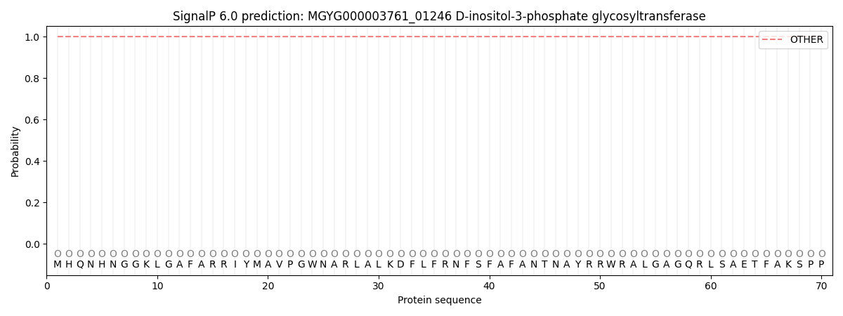
SignalP and Lipop Annotations help
This protein is predicted as OTHER
| Other | SP_Sec_SPI | LIPO_Sec_SPII | TAT_Tat_SPI | TATLIP_Sec_SPII | PILIN_Sec_SPIII |
|---|---|---|---|---|---|
| 1.000037 | 0.000003 | 0.000000 | 0.000000 | 0.000000 | 0.000000 |