You are browsing environment: HUMAN GUT
CAZyme Information: MGYG000003846_00137
You are here: Home > Sequence: MGYG000003846_00137
Basic Information |
Genomic context |
Full Sequence |
Enzyme annotations |
CAZy signature domains |
CDD domains |
CAZyme hits |
PDB hits |
Swiss-Prot hits |
SignalP and Lipop annotations |
TMHMM annotations
Basic Information help
| Species | ||||||||||||
|---|---|---|---|---|---|---|---|---|---|---|---|---|
| Lineage | Bacteria; Verrucomicrobiota; Verrucomicrobiae; Opitutales; CAG-312; CAG-312; | |||||||||||
| CAZyme ID | MGYG000003846_00137 | |||||||||||
| CAZy Family | GH10 | |||||||||||
| CAZyme Description | hypothetical protein | |||||||||||
| CAZyme Property |
|
|||||||||||
| Genome Property |
|
|||||||||||
| Gene Location | Start: 166352; End: 167944 Strand: - | |||||||||||
CAZyme Signature Domains help
| Family | Start | End | Evalue | family coverage |
|---|---|---|---|---|
| GH10 | 81 | 480 | 3.2e-42 | 0.9636963696369637 |
CDD Domains download full data without filtering help
| Cdd ID | Domain | E-Value | qStart | qEnd | sStart | sEnd | Domain Description |
|---|---|---|---|---|---|---|---|
| smart00633 | Glyco_10 | 1.13e-33 | 162 | 480 | 17 | 263 | Glycosyl hydrolase family 10. |
| pfam00331 | Glyco_hydro_10 | 1.99e-28 | 164 | 480 | 62 | 308 | Glycosyl hydrolase family 10. |
| COG3693 | XynA | 1.08e-25 | 158 | 486 | 79 | 343 | Endo-1,4-beta-xylanase, GH35 family [Carbohydrate transport and metabolism]. |
CAZyme Hits help
| Hit ID | E-Value | Query Start | Query End | Hit Start | Hit End |
|---|---|---|---|---|---|
| QGA28189.1 | 2.50e-165 | 26 | 530 | 19 | 504 |
| AHF92621.1 | 9.84e-163 | 33 | 520 | 16 | 481 |
| QQZ02681.1 | 1.06e-159 | 28 | 520 | 18 | 485 |
| AVM47074.1 | 2.91e-158 | 20 | 520 | 3 | 486 |
| AWI10666.1 | 2.12e-156 | 35 | 530 | 1 | 480 |
PDB Hits download full data without filtering help
| Hit ID | E-Value | Query Start | Query End | Hit Start | Hit End | Description |
|---|---|---|---|---|---|---|
| 3W24_A | 1.84e-15 | 163 | 483 | 68 | 326 | Thehigh-resolution crystal structure of TsXylA, intracellular xylanase from Thermoanaerobacterium saccharolyticum JW/SL-YS485 [Thermoanaerobacterium saccharolyticum JW/SL-YS485] |
| 3W27_A | 3.32e-15 | 163 | 483 | 68 | 326 | Thehigh-resolution crystal structure of TsXylA, intracellular xylanase from /Thermoanaerobacterium saccharolyticum JW/SL-YS485/: the complex of the E251A mutant with xylobiose [Thermoanaerobacterium saccharolyticum JW/SL-YS485],3W28_A The high-resolution crystal structure of TsXylA, intracellular xylanase from /Thermoanaerobacterium saccharolyticum JW/SL-YS485/: the complex of the E251A mutant with xylotriose [Thermoanaerobacterium saccharolyticum JW/SL-YS485],3W29_A The high-resolution crystal structure of TsXylA, intracellular xylanase from /Thermoanaerobacterium saccharolyticum JW/SL-YS485/: the complex of the E251A mutant with xylotetraose [Thermoanaerobacterium saccharolyticum JW/SL-YS485] |
| 3W25_A | 1.07e-14 | 163 | 483 | 68 | 326 | Thehigh-resolution crystal structure of TsXylA, intracellular xylanase from /Thermoanaerobacterium saccharolyticum JW/SL-YS485/: the complex of the E146A mutant with xylobiose [Thermoanaerobacterium saccharolyticum JW/SL-YS485],3W26_A The high-resolution crystal structure of TsXylA, intracellular xylanase from /Thermoanaerobacterium saccharolyticum JW/SL-YS485/: the complex of the E146A mutant with xylotriose [Thermoanaerobacterium saccharolyticum JW/SL-YS485] |
| 3RDK_A | 6.70e-13 | 163 | 480 | 64 | 335 | Proteincrystal structure of xylanase A1 of Paenibacillus sp. JDR-2 [Paenibacillus sp. JDR-2],3RDK_B Protein crystal structure of xylanase A1 of Paenibacillus sp. JDR-2 [Paenibacillus sp. JDR-2],3RO8_A Crystal structure of the catalytic domain of XynA1 from Paenibacillus sp. JDR-2 [Paenibacillus sp. JDR-2],3RO8_B Crystal structure of the catalytic domain of XynA1 from Paenibacillus sp. JDR-2 [Paenibacillus sp. JDR-2],3RO8_C Crystal structure of the catalytic domain of XynA1 from Paenibacillus sp. JDR-2 [Paenibacillus sp. JDR-2],3RO8_D Crystal structure of the catalytic domain of XynA1 from Paenibacillus sp. JDR-2 [Paenibacillus sp. JDR-2],3RO8_E Crystal structure of the catalytic domain of XynA1 from Paenibacillus sp. JDR-2 [Paenibacillus sp. JDR-2],3RO8_F Crystal structure of the catalytic domain of XynA1 from Paenibacillus sp. JDR-2 [Paenibacillus sp. JDR-2],3RO8_G Crystal structure of the catalytic domain of XynA1 from Paenibacillus sp. JDR-2 [Paenibacillus sp. JDR-2],3RO8_H Crystal structure of the catalytic domain of XynA1 from Paenibacillus sp. JDR-2 [Paenibacillus sp. JDR-2],4E4P_A Second native structure of Xylanase A1 from Paenibacillus sp. JDR-2 [Paenibacillus sp. JDR-2],4E4P_B Second native structure of Xylanase A1 from Paenibacillus sp. JDR-2 [Paenibacillus sp. JDR-2] |
| 6LPS_A | 4.11e-12 | 158 | 480 | 64 | 349 | ChainA, Beta-xylanase [Halalkalibacterium halodurans] |
Swiss-Prot Hits download full data without filtering help
| Hit ID | E-Value | Query Start | Query End | Hit Start | Hit End | Description |
|---|---|---|---|---|---|---|
| C6CRV0 | 1.36e-11 | 163 | 480 | 577 | 848 | Endo-1,4-beta-xylanase A OS=Paenibacillus sp. (strain JDR-2) OX=324057 GN=xynA1 PE=1 SV=1 |
| A1CHQ0 | 5.56e-11 | 163 | 480 | 90 | 313 | Probable endo-1,4-beta-xylanase C OS=Aspergillus clavatus (strain ATCC 1007 / CBS 513.65 / DSM 816 / NCTC 3887 / NRRL 1 / QM 1276 / 107) OX=344612 GN=xlnC PE=2 SV=1 |
| P07528 | 1.61e-10 | 148 | 361 | 95 | 273 | Endo-1,4-beta-xylanase A OS=Alkalihalobacillus halodurans (strain ATCC BAA-125 / DSM 18197 / FERM 7344 / JCM 9153 / C-125) OX=272558 GN=xynA PE=1 SV=1 |
| P40942 | 6.46e-10 | 163 | 480 | 104 | 379 | Thermostable celloxylanase OS=Thermoclostridium stercorarium OX=1510 GN=xynB PE=1 SV=1 |
| P23556 | 5.10e-09 | 151 | 480 | 63 | 339 | Endo-1,4-beta-xylanase A OS=Caldicellulosiruptor saccharolyticus OX=44001 GN=xynA PE=1 SV=1 |
SignalP and Lipop Annotations help
This protein is predicted as SP
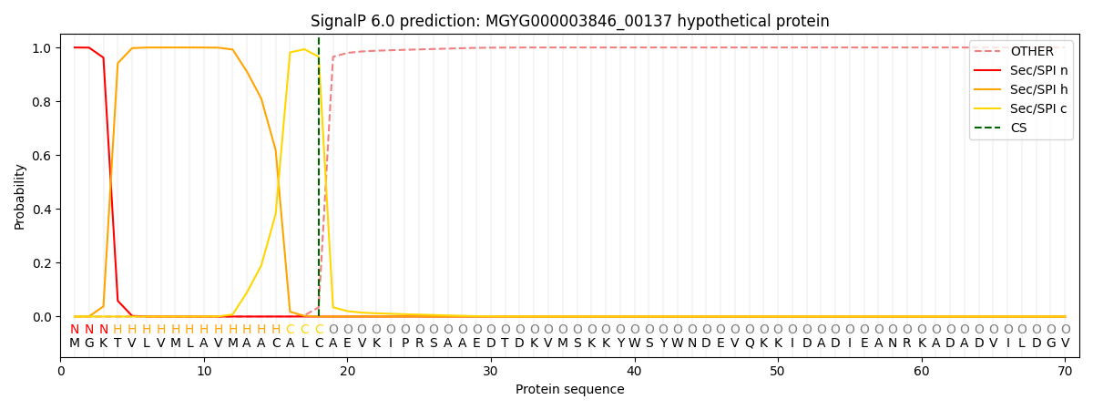
| Other | SP_Sec_SPI | LIPO_Sec_SPII | TAT_Tat_SPI | TATLIP_Sec_SPII | PILIN_Sec_SPIII |
|---|---|---|---|---|---|
| 0.000301 | 0.998645 | 0.000538 | 0.000180 | 0.000153 | 0.000146 |