You are browsing environment: HUMAN GUT
CAZyme Information: MGYG000003931_01135
You are here: Home > Sequence: MGYG000003931_01135
Basic Information |
Genomic context |
Full Sequence |
Enzyme annotations |
CAZy signature domains |
CDD domains |
CAZyme hits |
PDB hits |
Swiss-Prot hits |
SignalP and Lipop annotations |
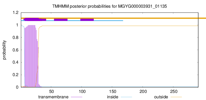
TMHMM annotations
Basic Information help
| Species | ||||||||||||
|---|---|---|---|---|---|---|---|---|---|---|---|---|
| Lineage | Bacteria; Firmicutes_A; Clostridia_A; Christensenellales; CAG-552; UMGS1880; | |||||||||||
| CAZyme ID | MGYG000003931_01135 | |||||||||||
| CAZy Family | GH16 | |||||||||||
| CAZyme Description | hypothetical protein | |||||||||||
| CAZyme Property |
|
|||||||||||
| Genome Property |
|
|||||||||||
| Gene Location | Start: 3583; End: 4458 Strand: - | |||||||||||
CAZyme Signature Domains help
| Family | Start | End | Evalue | family coverage |
|---|---|---|---|---|
| GH16 | 86 | 285 | 2.5e-65 | 0.9514563106796117 |
CDD Domains download full data without filtering help
| Cdd ID | Domain | E-Value | qStart | qEnd | sStart | sEnd | Domain Description |
|---|---|---|---|---|---|---|---|
| cd02175 | GH16_lichenase | 5.00e-82 | 82 | 289 | 7 | 212 | lichenase, member of glycosyl hydrolase family 16. Lichenase, also known as 1,3-1,4-beta-glucanase, is a member of glycosyl hydrolase family 16, that specifically cleaves 1,4-beta-D-glucosidic bonds in mixed-linked beta glucans that also contain 1,3-beta-D-glucosidic linkages. Natural substrates of beta-glucanase are beta-glucans from grain endosperm cell walls or lichenan from the Islandic moss, Cetraria islandica. This protein is found not only in bacteria but also in anaerobic fungi. This domain includes two seven-stranded antiparallel beta-sheets that are adjacent to one another forming a compact, jellyroll beta-sandwich structure. |
| pfam00722 | Glyco_hydro_16 | 5.40e-45 | 104 | 286 | 1 | 168 | Glycosyl hydrolases family 16. |
| cd00413 | Glyco_hydrolase_16 | 6.01e-44 | 76 | 285 | 2 | 207 | glycosyl hydrolase family 16. The O-Glycosyl hydrolases are a widespread group of enzymes that hydrolyse the glycosidic bond between two or more carbohydrates, or between a carbohydrate and a non-carbohydrate moiety. A glycosyl hydrolase classification system based on sequence similarity has led to the definition of more than 95 different families inlcuding glycosyl hydrolase family 16. Family 16 includes lichenase, xyloglucan endotransglycosylase (XET), beta-agarase, kappa-carrageenase, endo-beta-1,3-glucanase, endo-beta-1,3-1,4-glucanase, and endo-beta-galactosidase, all of which have a conserved jelly roll fold with a deep active site channel harboring the catalytic residues. |
| COG2273 | BglS | 7.03e-39 | 90 | 289 | 56 | 264 | Beta-glucanase, GH16 family [Carbohydrate transport and metabolism]. |
| cd02176 | GH16_XET | 1.76e-25 | 137 | 276 | 35 | 185 | Xyloglucan endotransglycosylase, member of glycosyl hydrolase family 16. Xyloglucan endotransglycosylases (XETs) cleave and religate xyloglucan polymers in plant cell walls via a transglycosylation mechanism. Xyloglucan is a soluble hemicellulose with a backbone of beta-1,4-linked glucose units, partially substituted with alpha-1,6-linked xylopyranose branches. It binds noncovalently to cellulose, cross-linking the adjacent cellulose microfibrils, giving it a key structural role as a matrix polymer. Therefore, XET plays an important role in all plant processes that require cell wall remodeling. |
CAZyme Hits help
| Hit ID | E-Value | Query Start | Query End | Hit Start | Hit End |
|---|---|---|---|---|---|
| ADD61790.1 | 2.50e-69 | 45 | 290 | 26 | 256 |
| CCO06214.1 | 1.33e-68 | 52 | 289 | 21 | 253 |
| AIQ43119.1 | 2.45e-65 | 88 | 287 | 44 | 237 |
| QCT01047.1 | 3.03e-65 | 88 | 285 | 62 | 253 |
| AIQ31818.1 | 9.83e-65 | 88 | 287 | 44 | 237 |
PDB Hits download full data without filtering help
| Hit ID | E-Value | Query Start | Query End | Hit Start | Hit End | Description |
|---|---|---|---|---|---|---|
| 1MAC_A | 1.19e-65 | 88 | 285 | 16 | 207 | CrystalStructure And Site-Directed Mutagenesis Of Bacillus Macerans Endo-1,3-1,4-Beta-Glucanase [Paenibacillus macerans],1MAC_B Crystal Structure And Site-Directed Mutagenesis Of Bacillus Macerans Endo-1,3-1,4-Beta-Glucanase [Paenibacillus macerans] |
| 1BYH_A | 1.27e-65 | 84 | 285 | 9 | 209 | MOLECULARAND ACTIVE-SITE STRUCTURE OF A BACILLUS (1-3,1-4)-BETA-GLUCANASE [synthetic construct],1GLH_A Cation Binding To A Bacillus (1,3-1,4)-Beta-Glucanase. Geometry, Affinity And Effect On Protein Stability [Paenibacillus macerans],2AYH_A Crystal And Molecular Structure At 1.6 Angstroms Resolution Of The Hybrid Bacillus Endo-1,3-1,4-Beta-D-Glucan 4- Glucanohydrolase H(A16-M) [hybrid] |
| 1U0A_A | 1.02e-64 | 84 | 285 | 9 | 209 | ChainA, Beta-glucanase [Paenibacillus macerans],1U0A_B Chain B, Beta-glucanase [Paenibacillus macerans],1U0A_C Chain C, Beta-glucanase [Paenibacillus macerans],1U0A_D Chain D, Beta-glucanase [Paenibacillus macerans] |
| 1GBG_A | 9.33e-63 | 84 | 285 | 9 | 209 | BacillusLicheniformis Beta-Glucanase [Bacillus licheniformis] |
| 3O5S_A | 1.97e-62 | 74 | 285 | 21 | 233 | CrystalStructure of the endo-beta-1,3-1,4 glucanase from Bacillus subtilis (strain 168) [Bacillus subtilis] |
Swiss-Prot Hits download full data without filtering help
| Hit ID | E-Value | Query Start | Query End | Hit Start | Hit End | Description |
|---|---|---|---|---|---|---|
| P23904 | 1.44e-64 | 88 | 285 | 41 | 232 | Beta-glucanase OS=Paenibacillus macerans OX=44252 PE=1 SV=2 |
| P45797 | 8.41e-64 | 77 | 285 | 20 | 233 | Beta-glucanase OS=Paenibacillus polymyxa OX=1406 GN=gluB PE=3 SV=1 |
| O14412 | 1.48e-63 | 74 | 289 | 39 | 242 | Beta-glucanase OS=Orpinomyces sp. (strain PC-2) OX=50059 GN=licA PE=1 SV=1 |
| P07980 | 2.78e-62 | 71 | 285 | 19 | 234 | Beta-glucanase OS=Bacillus amyloliquefaciens OX=1390 GN=bglA PE=3 SV=1 |
| P04957 | 2.44e-61 | 84 | 285 | 37 | 237 | Beta-glucanase OS=Bacillus subtilis (strain 168) OX=224308 GN=bglS PE=1 SV=2 |
SignalP and Lipop Annotations help
This protein is predicted as LIPO
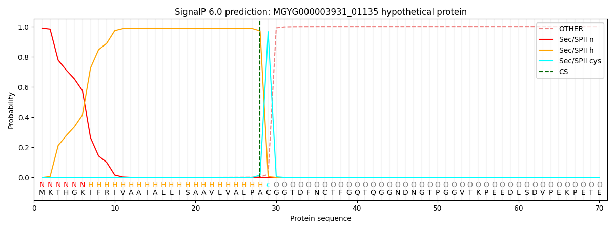
| Other | SP_Sec_SPI | LIPO_Sec_SPII | TAT_Tat_SPI | TATLIP_Sec_SPII | PILIN_Sec_SPIII |
|---|---|---|---|---|---|
| 0.000028 | 0.009144 | 0.990821 | 0.000003 | 0.000006 | 0.000005 |