You are browsing environment: HUMAN GUT
CAZyme Information: MGYG000003961_01415
You are here: Home > Sequence: MGYG000003961_01415
Basic Information |
Genomic context |
Full Sequence |
Enzyme annotations |
CAZy signature domains |
CDD domains |
CAZyme hits |
PDB hits |
Swiss-Prot hits |
SignalP and Lipop annotations |
TMHMM annotations
Basic Information help
| Species | RUG472 sp900545265 | |||||||||||
|---|---|---|---|---|---|---|---|---|---|---|---|---|
| Lineage | Bacteria; Firmicutes_A; Clostridia_A; Christensenellales; CAG-138; RUG472; RUG472 sp900545265 | |||||||||||
| CAZyme ID | MGYG000003961_01415 | |||||||||||
| CAZy Family | GH13 | |||||||||||
| CAZyme Description | 1,4-alpha-glucan branching enzyme GlgB | |||||||||||
| CAZyme Property |
|
|||||||||||
| Genome Property |
|
|||||||||||
| Gene Location | Start: 8055; End: 9743 Strand: + | |||||||||||
CAZyme Signature Domains help
| Family | Start | End | Evalue | family coverage |
|---|---|---|---|---|
| GH13 | 103 | 403 | 9.1e-142 | 0.9966777408637874 |
CDD Domains download full data without filtering help
| Cdd ID | Domain | E-Value | qStart | qEnd | sStart | sEnd | Domain Description |
|---|---|---|---|---|---|---|---|
| PRK05402 | PRK05402 | 0.0 | 1 | 546 | 161 | 724 | 1,4-alpha-glucan branching protein GlgB. |
| cd11322 | AmyAc_Glg_BE | 0.0 | 48 | 439 | 4 | 402 | Alpha amylase catalytic domain found in the Glycogen branching enzyme (also called 1,4-alpha-glucan branching enzyme). The glycogen branching enzyme catalyzes the third step of glycogen biosynthesis by the cleavage of an alpha-(1,4)-glucosidic linkage and the formation a new alpha-(1,6)-branch by subsequent transfer of cleaved oligosaccharide. They are part of a group called branching enzymes which catalyze the formation of alpha-1,6 branch points in either glycogen or starch. This group includes proteins from bacteria, eukaryotes, and archaea. The Alpha-amylase family comprises the largest family of glycoside hydrolases (GH), with the majority of enzymes acting on starch, glycogen, and related oligo- and polysaccharides. These proteins catalyze the transformation of alpha-1,4 and alpha-1,6 glucosidic linkages with retention of the anomeric center. The protein is described as having 3 domains: A, B, C. A is a (beta/alpha) 8-barrel; B is a loop between the beta 3 strand and alpha 3 helix of A; C is the C-terminal extension characterized by a Greek key. The majority of the enzymes have an active site cleft found between domains A and B where a triad of catalytic residues (Asp, Glu and Asp) performs catalysis. Other members of this family have lost the catalytic activity as in the case of the human 4F2hc, or only have 2 residues that serve as the catalytic nucleophile and the acid/base, such as Thermus A4 beta-galactosidase with 2 Glu residues (GH42) and human alpha-galactosidase with 2 Asp residues (GH31). The family members are quite extensive and include: alpha amylase, maltosyltransferase, cyclodextrin glycotransferase, maltogenic amylase, neopullulanase, isoamylase, 1,4-alpha-D-glucan maltotetrahydrolase, 4-alpha-glucotransferase, oligo-1,6-glucosidase, amylosucrase, sucrose phosphorylase, and amylomaltase. |
| PRK14705 | PRK14705 | 0.0 | 7 | 543 | 675 | 1220 | glycogen branching enzyme; Provisional |
| PRK12313 | PRK12313 | 0.0 | 1 | 551 | 68 | 633 | 1,4-alpha-glucan branching protein GlgB. |
| TIGR01515 | branching_enzym | 0.0 | 1 | 544 | 58 | 618 | alpha-1,4-glucan:alpha-1,4-glucan 6-glycosyltransferase. This model describes the glycogen branching enzymes which are responsible for the transfer of chains of approx. 7 alpha(1--4)-linked glucosyl residues to other similar chains (in new alpha(1--6) linkages) in the biosynthesis of glycogen. This enzyme is a member of the broader amylase family of starch hydrolases which fold as (beta/alpha)8 barrels, the so-called TIM-barrel structure. All of the sequences comprising the seed of this model have been experimentally characterized. This model encompasses both bacterial and eukaryotic species. No archaea have this enzyme, although Aquifex aolicus does. Two species, Bacillus thuringiensis and Clostridium perfringens have two sequences each which are annotated as amylases. These annotations are aparrently in error. GP|18143720 from C. perfringens, for instance, contains the note "674 aa, similar to gp:A14658_1 amylase (1,4-alpha-glucan branching enzyme (EC 2.4.1.18) ) from Bacillus thuringiensis (648 aa); 51.1% identity in 632 aa overlap." A branching enzyme from Porphyromonas gingivales, OMNI|PG1793, appears to be more closely related to the eukaryotic species (across a deep phylogenetic split) and may represent an instance of lateral transfer from this species' host. A sequence from Arabidopsis thaliana, GP|9294564, scores just above trusted, but appears either to contain corrupt sequence or, more likely, to be a pseudogene as some of the conserved catalytic residues common to the alpha amylase family are not conserved here. [Energy metabolism, Biosynthesis and degradation of polysaccharides] |
CAZyme Hits help
| Hit ID | E-Value | Query Start | Query End | Hit Start | Hit End |
|---|---|---|---|---|---|
| QCX33360.1 | 9.99e-209 | 7 | 545 | 68 | 621 |
| CBL01646.1 | 1.51e-208 | 1 | 560 | 73 | 653 |
| AXB29737.1 | 2.69e-207 | 1 | 560 | 73 | 653 |
| CBK98932.1 | 8.41e-204 | 6 | 545 | 79 | 633 |
| ATL90043.1 | 1.67e-203 | 6 | 545 | 79 | 633 |
PDB Hits download full data without filtering help
| Hit ID | E-Value | Query Start | Query End | Hit Start | Hit End | Description |
|---|---|---|---|---|---|---|
| 6JOY_A | 6.67e-177 | 1 | 548 | 61 | 621 | TheX-ray Crystallographic Structure of Branching Enzyme from Rhodothermus obamensis STB05 [Rhodothermus marinus] |
| 4LPC_A | 2.70e-171 | 3 | 546 | 53 | 610 | CrystalStructure of E.Coli Branching Enzyme in complex with maltoheptaose [Escherichia coli],4LPC_B Crystal Structure of E.Coli Branching Enzyme in complex with maltoheptaose [Escherichia coli],4LPC_C Crystal Structure of E.Coli Branching Enzyme in complex with maltoheptaose [Escherichia coli],4LPC_D Crystal Structure of E.Coli Branching Enzyme in complex with maltoheptaose [Escherichia coli],4LQ1_A Crystal Structure of E.Coli Branching Enzyme in complex with maltohexaose [Escherichia coli],4LQ1_B Crystal Structure of E.Coli Branching Enzyme in complex with maltohexaose [Escherichia coli],4LQ1_C Crystal Structure of E.Coli Branching Enzyme in complex with maltohexaose [Escherichia coli],4LQ1_D Crystal Structure of E.Coli Branching Enzyme in complex with maltohexaose [Escherichia coli],5E6Y_A Crystal structure of E.Coli branching enzyme in complex with alpha cyclodextrin [Escherichia coli O139:H28 str. E24377A],5E6Y_B Crystal structure of E.Coli branching enzyme in complex with alpha cyclodextrin [Escherichia coli O139:H28 str. E24377A],5E6Y_C Crystal structure of E.Coli branching enzyme in complex with alpha cyclodextrin [Escherichia coli O139:H28 str. E24377A],5E6Y_D Crystal structure of E.Coli branching enzyme in complex with alpha cyclodextrin [Escherichia coli O139:H28 str. E24377A],5E6Z_A Crystal structure of Ecoli Branching Enzyme with beta cyclodextrin [Escherichia coli O139:H28 str. E24377A],5E6Z_B Crystal structure of Ecoli Branching Enzyme with beta cyclodextrin [Escherichia coli O139:H28 str. E24377A],5E6Z_C Crystal structure of Ecoli Branching Enzyme with beta cyclodextrin [Escherichia coli O139:H28 str. E24377A],5E6Z_D Crystal structure of Ecoli Branching Enzyme with beta cyclodextrin [Escherichia coli O139:H28 str. E24377A],5E70_A Crystal structure of Ecoli Branching Enzyme with gamma cyclodextrin [Escherichia coli O139:H28 str. E24377A],5E70_B Crystal structure of Ecoli Branching Enzyme with gamma cyclodextrin [Escherichia coli O139:H28 str. E24377A],5E70_C Crystal structure of Ecoli Branching Enzyme with gamma cyclodextrin [Escherichia coli O139:H28 str. E24377A],5E70_D Crystal structure of Ecoli Branching Enzyme with gamma cyclodextrin [Escherichia coli O139:H28 str. E24377A] |
| 1M7X_A | 3.18e-171 | 3 | 546 | 58 | 615 | TheX-ray Crystallographic Structure of Branching Enzyme [Escherichia coli],1M7X_B The X-ray Crystallographic Structure of Branching Enzyme [Escherichia coli],1M7X_C The X-ray Crystallographic Structure of Branching Enzyme [Escherichia coli],1M7X_D The X-ray Crystallographic Structure of Branching Enzyme [Escherichia coli] |
| 3K1D_A | 6.13e-170 | 5 | 537 | 171 | 713 | Crystalstructure of glycogen branching enzyme synonym: 1,4-alpha-D-glucan:1,4-alpha-D-GLUCAN 6-glucosyl-transferase from mycobacterium tuberculosis H37RV [Mycobacterium tuberculosis H37Rv] |
| 5GR1_A | 6.55e-167 | 1 | 546 | 190 | 774 | Crystalstructure of branching enzyme Y500A/D501A mutant from Cyanothece sp. ATCC 51142 in complex with maltoheptaose [Crocosphaera subtropica ATCC 51142],5GR6_A Crystal structure of branching enzyme Y500A/D501A double mutant from Cyanothece sp. ATCC 51142 [Crocosphaera subtropica ATCC 51142] |
Swiss-Prot Hits download full data without filtering help
| Hit ID | E-Value | Query Start | Query End | Hit Start | Hit End | Description |
|---|---|---|---|---|---|---|
| B3PGN4 | 6.22e-198 | 3 | 545 | 179 | 736 | 1,4-alpha-glucan branching enzyme GlgB OS=Cellvibrio japonicus (strain Ueda107) OX=498211 GN=glgB PE=3 SV=1 |
| Q1H1K2 | 4.22e-190 | 7 | 544 | 170 | 723 | 1,4-alpha-glucan branching enzyme GlgB OS=Methylobacillus flagellatus (strain KT / ATCC 51484 / DSM 6875) OX=265072 GN=glgB PE=3 SV=1 |
| Q3JCN0 | 3.62e-189 | 7 | 546 | 179 | 733 | 1,4-alpha-glucan branching enzyme GlgB OS=Nitrosococcus oceani (strain ATCC 19707 / BCRC 17464 / JCM 30415 / NCIMB 11848 / C-107) OX=323261 GN=glgB PE=3 SV=1 |
| B8CVY1 | 6.57e-187 | 1 | 544 | 67 | 626 | 1,4-alpha-glucan branching enzyme GlgB OS=Halothermothrix orenii (strain H 168 / OCM 544 / DSM 9562) OX=373903 GN=glgB PE=3 SV=1 |
| Q5Z0W8 | 3.20e-186 | 7 | 542 | 195 | 738 | 1,4-alpha-glucan branching enzyme GlgB OS=Nocardia farcinica (strain IFM 10152) OX=247156 GN=glgB PE=3 SV=1 |
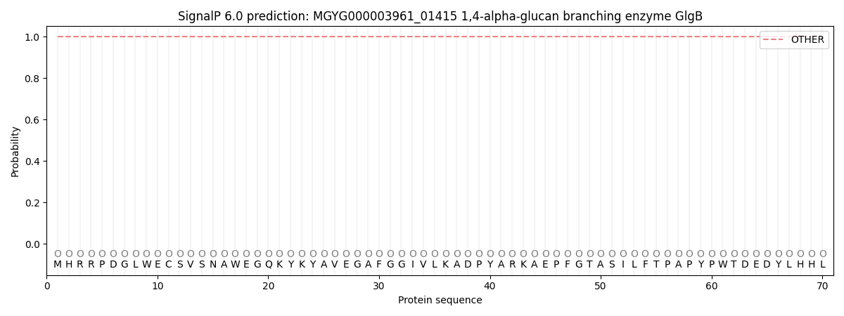
SignalP and Lipop Annotations help
This protein is predicted as OTHER
| Other | SP_Sec_SPI | LIPO_Sec_SPII | TAT_Tat_SPI | TATLIP_Sec_SPII | PILIN_Sec_SPIII |
|---|---|---|---|---|---|
| 1.000065 | 0.000001 | 0.000000 | 0.000000 | 0.000000 | 0.000000 |