You are browsing environment: HUMAN GUT
CAZyme Information: MGYG000004044_00316
You are here: Home > Sequence: MGYG000004044_00316
Basic Information |
Genomic context |
Full Sequence |
Enzyme annotations |
CAZy signature domains |
CDD domains |
CAZyme hits |
PDB hits |
Swiss-Prot hits |
SignalP and Lipop annotations |
TMHMM annotations
Basic Information help
| Species | UMGS1633 sp900553645 | |||||||||||
|---|---|---|---|---|---|---|---|---|---|---|---|---|
| Lineage | Bacteria; Firmicutes_A; Clostridia_A; Christensenellales; CAG-74; UMGS1633; UMGS1633 sp900553645 | |||||||||||
| CAZyme ID | MGYG000004044_00316 | |||||||||||
| CAZy Family | GH39 | |||||||||||
| CAZyme Description | hypothetical protein | |||||||||||
| CAZyme Property |
|
|||||||||||
| Genome Property |
|
|||||||||||
| Gene Location | Start: 18199; End: 19527 Strand: - | |||||||||||
CAZyme Signature Domains help
| Family | Start | End | Evalue | family coverage |
|---|---|---|---|---|
| GH39 | 68 | 387 | 3.4e-33 | 0.7378190255220418 |
CDD Domains download full data without filtering help
| Cdd ID | Domain | E-Value | qStart | qEnd | sStart | sEnd | Domain Description |
|---|---|---|---|---|---|---|---|
| pfam01229 | Glyco_hydro_39 | 1.84e-15 | 3 | 388 | 2 | 391 | Glycosyl hydrolases family 39. |
| cd21510 | agarase_cat | 2.58e-09 | 120 | 254 | 73 | 187 | alpha-beta barrel catalytic domain of agarase, such as GH86-like endo-acting agarases identified in non-marine organisms. Typically, agarases (E.C. 3.2.1.81) are found in ocean-dwelling bacteria since agarose is a principle component of red algae cell wall polysaccharides. Agarose is a linear polymer of alternating D-galactose and 3,6-anhydro-L-galactopyranose. Endo-acting agarases, such as glycoside hydrolase 16 (GH16) and GH86 hydrolyze internal beta-1,4 linkages. GH86-like endo-acting agarase of this protein family has been identified in the human intestinal bacterium Bacteroides uniformis. This acquired metabolic pathway, as demonstrated by the prevalence of agar-specific genetic cluster called polysaccharide utilization loci (PULs), varies considerably between human populations, being much more prevalent in a Japanese sample than in North America, European, or Chinese samples. Agarase activity was also identified in the non-marine bacterium Cellvibrio sp. |
| pfam12891 | Glyco_hydro_44 | 8.27e-05 | 120 | 207 | 101 | 185 | Glycoside hydrolase family 44. This is a family of bacterial glycoside hydrolases formerly known as cellulase family J, and now known as Cel44A. It is one of the major enzymatic components of the cellulosome of Clostridium thermocellum strain F1 and of many other Firmicutes. |
CAZyme Hits help
| Hit ID | E-Value | Query Start | Query End | Hit Start | Hit End |
|---|---|---|---|---|---|
| AVM69820.1 | 5.06e-137 | 4 | 440 | 3 | 446 |
| QJD85864.1 | 6.03e-76 | 1 | 430 | 1 | 435 |
| QJD82796.1 | 3.33e-65 | 9 | 386 | 8 | 378 |
| QTH43246.1 | 7.37e-63 | 9 | 386 | 8 | 378 |
| AVM46100.1 | 1.97e-57 | 7 | 415 | 7 | 407 |
PDB Hits download full data without filtering help
| Hit ID | E-Value | Query Start | Query End | Hit Start | Hit End | Description |
|---|---|---|---|---|---|---|
| 5Z3K_A | 8.76e-19 | 1 | 287 | 6 | 266 | Crystalstructure of glucosidase from Croceicoccus marinus at 1.8 Angstrom resolution [Croceicoccus marinus],5Z3K_B Crystal structure of glucosidase from Croceicoccus marinus at 1.8 Angstrom resolution [Croceicoccus marinus] |
| 1W91_A | 2.29e-15 | 51 | 266 | 47 | 242 | crystalstructure of 1,4-BETA-D-XYLAN XYLOHYDROLASE solve using anomalous signal from Seleniomethionine [synthetic construct],1W91_B crystal structure of 1,4-BETA-D-XYLAN XYLOHYDROLASE solve using anomalous signal from Seleniomethionine [synthetic construct],1W91_C crystal structure of 1,4-BETA-D-XYLAN XYLOHYDROLASE solve using anomalous signal from Seleniomethionine [synthetic construct],1W91_D crystal structure of 1,4-BETA-D-XYLAN XYLOHYDROLASE solve using anomalous signal from Seleniomethionine [synthetic construct],1W91_E crystal structure of 1,4-BETA-D-XYLAN XYLOHYDROLASE solve using anomalous signal from Seleniomethionine [synthetic construct],1W91_F crystal structure of 1,4-BETA-D-XYLAN XYLOHYDROLASE solve using anomalous signal from Seleniomethionine [synthetic construct],1W91_G crystal structure of 1,4-BETA-D-XYLAN XYLOHYDROLASE solve using anomalous signal from Seleniomethionine [synthetic construct],1W91_H crystal structure of 1,4-BETA-D-XYLAN XYLOHYDROLASE solve using anomalous signal from Seleniomethionine [synthetic construct] |
| 2BS9_A | 2.29e-15 | 51 | 266 | 47 | 242 | Nativecrystal structure of a GH39 beta-xylosidase XynB1 from Geobacillus stearothermophilus [Geobacillus stearothermophilus],2BS9_B Native crystal structure of a GH39 beta-xylosidase XynB1 from Geobacillus stearothermophilus [Geobacillus stearothermophilus],2BS9_C Native crystal structure of a GH39 beta-xylosidase XynB1 from Geobacillus stearothermophilus [Geobacillus stearothermophilus],2BS9_D Native crystal structure of a GH39 beta-xylosidase XynB1 from Geobacillus stearothermophilus [Geobacillus stearothermophilus],2BS9_E Native crystal structure of a GH39 beta-xylosidase XynB1 from Geobacillus stearothermophilus [Geobacillus stearothermophilus],2BS9_F Native crystal structure of a GH39 beta-xylosidase XynB1 from Geobacillus stearothermophilus [Geobacillus stearothermophilus],2BS9_G Native crystal structure of a GH39 beta-xylosidase XynB1 from Geobacillus stearothermophilus [Geobacillus stearothermophilus],2BS9_H Native crystal structure of a GH39 beta-xylosidase XynB1 from Geobacillus stearothermophilus [Geobacillus stearothermophilus] |
| 2BFG_A | 5.40e-15 | 51 | 266 | 47 | 242 | crystalstructure of beta-xylosidase (fam GH39) in complex with dinitrophenyl-beta-xyloside and covalently bound xyloside [Geobacillus stearothermophilus],2BFG_B crystal structure of beta-xylosidase (fam GH39) in complex with dinitrophenyl-beta-xyloside and covalently bound xyloside [Geobacillus stearothermophilus],2BFG_C crystal structure of beta-xylosidase (fam GH39) in complex with dinitrophenyl-beta-xyloside and covalently bound xyloside [Geobacillus stearothermophilus],2BFG_D crystal structure of beta-xylosidase (fam GH39) in complex with dinitrophenyl-beta-xyloside and covalently bound xyloside [Geobacillus stearothermophilus],2BFG_E crystal structure of beta-xylosidase (fam GH39) in complex with dinitrophenyl-beta-xyloside and covalently bound xyloside [Geobacillus stearothermophilus],2BFG_F crystal structure of beta-xylosidase (fam GH39) in complex with dinitrophenyl-beta-xyloside and covalently bound xyloside [Geobacillus stearothermophilus],2BFG_G crystal structure of beta-xylosidase (fam GH39) in complex with dinitrophenyl-beta-xyloside and covalently bound xyloside [Geobacillus stearothermophilus],2BFG_H crystal structure of beta-xylosidase (fam GH39) in complex with dinitrophenyl-beta-xyloside and covalently bound xyloside [Geobacillus stearothermophilus] |
| 1PX8_A | 1.22e-12 | 79 | 258 | 77 | 234 | Crystalstructure of beta-D-xylosidase from Thermoanaerobacterium saccharolyticum, a family 39 glycoside hydrolase [Thermoanaerobacterium saccharolyticum],1PX8_B Crystal structure of beta-D-xylosidase from Thermoanaerobacterium saccharolyticum, a family 39 glycoside hydrolase [Thermoanaerobacterium saccharolyticum],1UHV_A Crystal structure of beta-D-xylosidase from Thermoanaerobacterium saccharolyticum, a family 39 glycoside hydrolase [Thermoanaerobacterium saccharolyticum],1UHV_B Crystal structure of beta-D-xylosidase from Thermoanaerobacterium saccharolyticum, a family 39 glycoside hydrolase [Thermoanaerobacterium saccharolyticum],1UHV_C Crystal structure of beta-D-xylosidase from Thermoanaerobacterium saccharolyticum, a family 39 glycoside hydrolase [Thermoanaerobacterium saccharolyticum],1UHV_D Crystal structure of beta-D-xylosidase from Thermoanaerobacterium saccharolyticum, a family 39 glycoside hydrolase [Thermoanaerobacterium saccharolyticum] |
Swiss-Prot Hits download full data without filtering help
| Hit ID | E-Value | Query Start | Query End | Hit Start | Hit End | Description |
|---|---|---|---|---|---|---|
| Q9ZFM2 | 2.98e-15 | 51 | 352 | 47 | 307 | Beta-xylosidase OS=Geobacillus stearothermophilus OX=1422 GN=xynB PE=1 SV=1 |
| O30360 | 1.56e-11 | 79 | 258 | 77 | 234 | Beta-xylosidase OS=Thermoanaerobacterium saccharolyticum (strain DSM 8691 / JW/SL-YS485) OX=1094508 GN=xynB PE=3 SV=1 |
| P36906 | 8.58e-11 | 79 | 258 | 77 | 234 | Beta-xylosidase OS=Thermoanaerobacterium saccharolyticum OX=28896 GN=xynB PE=1 SV=1 |
SignalP and Lipop Annotations help
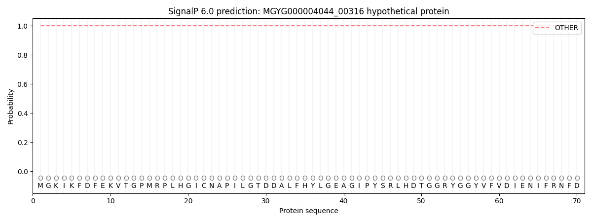
This protein is predicted as OTHER
| Other | SP_Sec_SPI | LIPO_Sec_SPII | TAT_Tat_SPI | TATLIP_Sec_SPII | PILIN_Sec_SPIII |
|---|---|---|---|---|---|
| 1.000041 | 0.000000 | 0.000000 | 0.000000 | 0.000000 | 0.000000 |