You are browsing environment: HUMAN GUT
CAZyme Information: MGYG000004094_00969
You are here: Home > Sequence: MGYG000004094_00969
Basic Information |
Genomic context |
Full Sequence |
Enzyme annotations |
CAZy signature domains |
CDD domains |
CAZyme hits |
PDB hits |
Swiss-Prot hits |
SignalP and Lipop annotations |
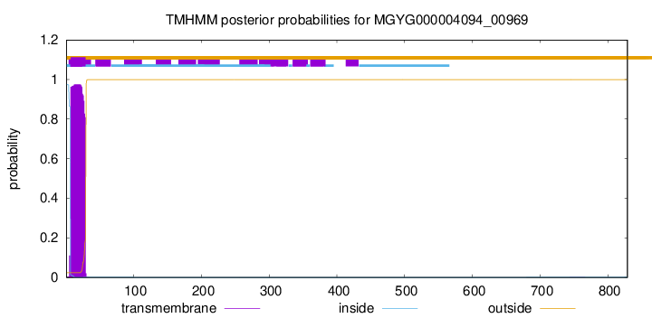
TMHMM annotations
Basic Information help
| Species | ||||||||||||
|---|---|---|---|---|---|---|---|---|---|---|---|---|
| Lineage | Bacteria; Bacteroidota; Bacteroidia; Bacteroidales; Bacteroidaceae; ; | |||||||||||
| CAZyme ID | MGYG000004094_00969 | |||||||||||
| CAZy Family | GH20 | |||||||||||
| CAZyme Description | hypothetical protein | |||||||||||
| CAZyme Property |
|
|||||||||||
| Genome Property |
|
|||||||||||
| Gene Location | Start: 109200; End: 111689 Strand: - | |||||||||||
CAZyme Signature Domains help
| Family | Start | End | Evalue | family coverage |
|---|---|---|---|---|
| GH20 | 138 | 432 | 2.1e-42 | 0.9554896142433235 |
CDD Domains download full data without filtering help
| Cdd ID | Domain | E-Value | qStart | qEnd | sStart | sEnd | Domain Description |
|---|---|---|---|---|---|---|---|
| cd06565 | GH20_GcnA-like | 4.84e-67 | 141 | 433 | 1 | 301 | Glycosyl hydrolase family 20 (GH20) catalytic domain of N-acetyl-beta-D-glucosaminidase (GcnA, also known as BhsA) and related proteins. GcnA is an exoglucosidase which cleaves N-acetyl-beta-D-galactosamine (NAG) and N-acetyl-beta-D-galactosamine residues from 4-methylumbelliferylated (4MU) substrates, as well as cleaving NAG from chito-oligosaccharides (i.e. NAG polymers). In contrast, sulfated forms of the substrate are unable to be cleaved and act instead as mild competitive inhibitors. Additionally, the enzyme is known to be poisoned by several first-row transition metals as well as by mercury. GcnA forms a homodimer with subunits comprised of three domains, an N-terminal zincin-like domain, this central catalytic GH20 domain, and a C-terminal alpha helical domain. The GH20 hexosaminidases are thought to act via a catalytic mechanism in which the catalytic nucleophile is not provided by solvent or the enzyme, but by the substrate itself. |
| pfam00728 | Glyco_hydro_20 | 3.37e-24 | 140 | 371 | 2 | 262 | Glycosyl hydrolase family 20, catalytic domain. This domain has a TIM barrel fold. |
| cd02742 | GH20_hexosaminidase | 4.23e-23 | 141 | 371 | 1 | 235 | Beta-N-acetylhexosaminidases of glycosyl hydrolase family 20 (GH20) catalyze the removal of beta-1,4-linked N-acetyl-D-hexosamine residues from the non-reducing ends of N-acetyl-beta-D-hexosaminides including N-acetylglucosides and N-acetylgalactosides. These enzymes are broadly distributed in microorganisms, plants and animals, and play roles in various key physiological and pathological processes. These processes include cell structural integrity, energy storage, cellular signaling, fertilization, pathogen defense, viral penetration, the development of carcinomas, inflammatory events and lysosomal storage disorders. The GH20 enzymes include the eukaryotic beta-N-acetylhexosaminidases A and B, the bacterial chitobiases, dispersin B, and lacto-N-biosidase. The GH20 hexosaminidases are thought to act via a catalytic mechanism in which the catalytic nucleophile is not provided by the solvent or the enzyme, but by the substrate itself. |
| COG3525 | Chb | 2.62e-22 | 86 | 391 | 204 | 559 | N-acetyl-beta-hexosaminidase [Carbohydrate transport and metabolism]. |
| cd06564 | GH20_DspB_LnbB-like | 4.74e-22 | 140 | 368 | 1 | 249 | Glycosyl hydrolase family 20 (GH20) catalytic domain of dispersin B (DspB), lacto-N-biosidase (LnbB) and related proteins. Dispersin B is a soluble beta-N-acetylglucosamidase found in bacteria that hydrolyzes the beta-1,6-linkages of PGA (poly-beta-(1,6)-N-acetylglucosamine), a major component of the extracellular polysaccharide matrix. Lacto-N-biosidase hydrolyzes lacto-N-biose (LNB) type I oligosaccharides at the nonreducing terminus to produce lacto-N-biose as part of the GNB/LNB (galacto-N-biose/lacto-N-biose I) degradation pathway. The lacto-N-biosidase from Bifidobacterium bifidum has this GH20 domain, a carbohydrate binding module 32, and a bacterial immunoglobulin-like domain 2, as well as a YSIRK signal peptide and a G5 membrane anchor at the N and C termini, respectively. The GH20 hexosaminidases are thought to act via a catalytic mechanism in which the catalytic nucleophile is not provided by solvent or the enzyme, but by the substrate itself. |
CAZyme Hits help
| Hit ID | E-Value | Query Start | Query End | Hit Start | Hit End |
|---|---|---|---|---|---|
| ANE51482.1 | 1.45e-162 | 51 | 825 | 76 | 848 |
| AXY74237.1 | 2.40e-155 | 65 | 825 | 89 | 843 |
| ACU05309.1 | 3.24e-153 | 81 | 828 | 103 | 838 |
| QRQ48164.1 | 1.20e-130 | 62 | 823 | 83 | 840 |
| QUT46231.1 | 2.36e-130 | 62 | 823 | 83 | 840 |
PDB Hits download full data without filtering help
| Hit ID | E-Value | Query Start | Query End | Hit Start | Hit End | Description |
|---|---|---|---|---|---|---|
| 6YHH_A | 1.45e-19 | 68 | 344 | 73 | 383 | X-rayStructure of Flavobacterium johnsoniae chitobiase (FjGH20) [Flavobacterium johnsoniae UW101],6YHH_B X-ray Structure of Flavobacterium johnsoniae chitobiase (FjGH20) [Flavobacterium johnsoniae UW101] |
| 3GH4_A | 1.02e-16 | 85 | 347 | 119 | 396 | Crystalstructure of beta-hexosaminidase from Paenibacillus sp. TS12 [Paenibacillus sp.],3GH5_A Crystal structure of beta-hexosaminidase from Paenibacillus sp. TS12 in complex with GlcNAc [Paenibacillus sp.],3GH7_A Crystal structure of beta-hexosaminidase from Paenibacillus sp. TS12 in complex with GalNAc [Paenibacillus sp.],3SUR_A Crystal structure of beta-hexosaminidase from Paenibacillus sp. TS12 in complex with NAG-thiazoline. [Paenibacillus sp. TS12],3SUS_A Crystal structure of beta-hexosaminidase from Paenibacillus sp. TS12 in complex with Gal-NAG-thiazoline [Paenibacillus sp. TS12],3SUT_A Crystal structure of beta-hexosaminidase from Paenibacillus sp. TS12 in complex with PUGNAc [Paenibacillus sp. TS12],3SUU_A Crystal structure of beta-hexosaminidase from Paenibacillus sp. TS12 in complex with Gal-PUGNAc [Paenibacillus sp. TS12],3SUV_A Crystal structure of beta-hexosaminidase from Paenibacillus sp. TS12 in complex with NHAc-DNJ [Paenibacillus sp. TS12],3SUW_A Crystal structure of beta-hexosaminidase from Paenibacillus sp. TS12 in complex with NHAc-CAS [Paenibacillus sp. TS12] |
| 6JQF_A | 6.23e-13 | 84 | 324 | 156 | 423 | Crystallizationanalysis of a beta-N-acetylhexosaminidase (Am2136) from Akkermansia muciniphila [Akkermansia muciniphila ATCC BAA-835] |
| 4PYS_A | 9.87e-13 | 29 | 353 | 34 | 370 | Thecrystal structure of beta-N-acetylhexosaminidase from Bacteroides fragilis NCTC 9343 [Bacteroides fragilis NCTC 9343],4PYS_B The crystal structure of beta-N-acetylhexosaminidase from Bacteroides fragilis NCTC 9343 [Bacteroides fragilis NCTC 9343] |
| 7DUP_A | 1.19e-10 | 88 | 344 | 79 | 389 | ChainA, Beta-N-acetylhexosaminidase [Bacteroides thetaiotaomicron],7DVA_A Chain A, Beta-N-acetylhexosaminidase [Bacteroides thetaiotaomicron],7DVA_B Chain B, Beta-N-acetylhexosaminidase [Bacteroides thetaiotaomicron] |
Swiss-Prot Hits download full data without filtering help
| Hit ID | E-Value | Query Start | Query End | Hit Start | Hit End | Description |
|---|---|---|---|---|---|---|
| P49008 | 2.35e-16 | 21 | 348 | 35 | 405 | Beta-hexosaminidase OS=Porphyromonas gingivalis (strain ATCC BAA-308 / W83) OX=242619 GN=nahA PE=3 SV=2 |
| B2UPR7 | 2.73e-15 | 84 | 344 | 178 | 464 | Beta-hexosaminidase Amuc_2136 OS=Akkermansia muciniphila (strain ATCC BAA-835 / DSM 22959 / JCM 33894 / BCRC 81048 / CCUG 64013 / CIP 107961 / Muc) OX=349741 GN=Amuc_2136 PE=1 SV=1 |
| Q9HGI3 | 1.52e-11 | 99 | 368 | 142 | 441 | Beta-hexosaminidase OS=Emericella nidulans OX=162425 GN=nagA PE=1 SV=1 |
| Q86M34 | 6.25e-09 | 87 | 344 | 132 | 417 | Beta-hexosaminidase subunit beta OS=Entamoeba histolytica OX=5759 GN=HEXB PE=1 SV=1 |
SignalP and Lipop Annotations help
This protein is predicted as SP
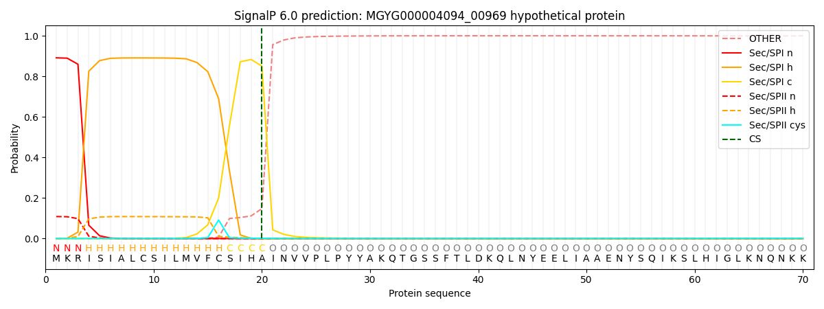
| Other | SP_Sec_SPI | LIPO_Sec_SPII | TAT_Tat_SPI | TATLIP_Sec_SPII | PILIN_Sec_SPIII |
|---|---|---|---|---|---|
| 0.001208 | 0.885228 | 0.112720 | 0.000292 | 0.000285 | 0.000262 |