You are browsing environment: HUMAN GUT
CAZyme Information: MGYG000004133_01690
You are here: Home > Sequence: MGYG000004133_01690
Basic Information |
Genomic context |
Full Sequence |
Enzyme annotations |
CAZy signature domains |
CDD domains |
CAZyme hits |
PDB hits |
Swiss-Prot hits |
SignalP and Lipop annotations |
TMHMM annotations
Basic Information help
| Species | ||||||||||||
|---|---|---|---|---|---|---|---|---|---|---|---|---|
| Lineage | Bacteria; Firmicutes_A; Clostridia; HGM11514; HGM11514; ; | |||||||||||
| CAZyme ID | MGYG000004133_01690 | |||||||||||
| CAZy Family | GH2 | |||||||||||
| CAZyme Description | Beta-glucuronidase | |||||||||||
| CAZyme Property |
|
|||||||||||
| Genome Property |
|
|||||||||||
| Gene Location | Start: 1981; End: 3744 Strand: + | |||||||||||
CAZyme Signature Domains help
| Family | Start | End | Evalue | family coverage |
|---|---|---|---|---|
| GH2 | 13 | 559 | 2.1e-94 | 0.5904255319148937 |
CDD Domains download full data without filtering help
| Cdd ID | Domain | E-Value | qStart | qEnd | sStart | sEnd | Domain Description |
|---|---|---|---|---|---|---|---|
| PRK10150 | PRK10150 | 9.36e-49 | 21 | 502 | 14 | 510 | beta-D-glucuronidase; Provisional |
| COG3250 | LacZ | 4.31e-36 | 12 | 454 | 6 | 444 | Beta-galactosidase/beta-glucuronidase [Carbohydrate transport and metabolism]. |
| PRK10340 | ebgA | 2.64e-18 | 51 | 438 | 99 | 472 | cryptic beta-D-galactosidase subunit alpha; Reviewed |
| pfam02837 | Glyco_hydro_2_N | 3.37e-18 | 22 | 179 | 4 | 169 | Glycosyl hydrolases family 2, sugar binding domain. This family contains beta-galactosidase, beta-mannosidase and beta-glucuronidase activities and has a jelly-roll fold. The domain binds the sugar moiety during the sugar-hydrolysis reaction. |
| PRK09525 | lacZ | 4.29e-09 | 75 | 501 | 124 | 539 | beta-galactosidase. |
CAZyme Hits help
| Hit ID | E-Value | Query Start | Query End | Hit Start | Hit End |
|---|---|---|---|---|---|
| ALS26546.1 | 4.27e-216 | 4 | 587 | 9 | 588 |
| AWP26385.1 | 9.87e-213 | 4 | 585 | 6 | 579 |
| ANA78861.1 | 1.40e-212 | 4 | 585 | 6 | 579 |
| AVV57223.1 | 1.40e-212 | 4 | 585 | 6 | 579 |
| QLG42947.1 | 3.12e-211 | 1 | 585 | 1 | 578 |
PDB Hits download full data without filtering help
| Hit ID | E-Value | Query Start | Query End | Hit Start | Hit End | Description |
|---|---|---|---|---|---|---|
| 7SF2_A | 7.89e-80 | 7 | 582 | 27 | 583 | ChainA, Glycosyl hydrolase family 2, sugar binding domain protein [Bacteroides cellulosilyticus DSM 14838],7SF2_B Chain B, Glycosyl hydrolase family 2, sugar binding domain protein [Bacteroides cellulosilyticus DSM 14838],7SF2_C Chain C, Glycosyl hydrolase family 2, sugar binding domain protein [Bacteroides cellulosilyticus DSM 14838],7SF2_D Chain D, Glycosyl hydrolase family 2, sugar binding domain protein [Bacteroides cellulosilyticus DSM 14838],7SF2_E Chain E, Glycosyl hydrolase family 2, sugar binding domain protein [Bacteroides cellulosilyticus DSM 14838],7SF2_F Chain F, Glycosyl hydrolase family 2, sugar binding domain protein [Bacteroides cellulosilyticus DSM 14838] |
| 4JHZ_A | 3.78e-32 | 22 | 502 | 17 | 508 | Structureof E. coli beta-Glucuronidase bound with a novel, potent inhibitor 2-[4-(1,3-benzodioxol-5-ylmethyl)piperazin-1-yl]-N-[(1S,2S,5S)-2,5-dimethoxycyclohexyl]acetamide [Escherichia coli K-12],4JHZ_B Structure of E. coli beta-Glucuronidase bound with a novel, potent inhibitor 2-[4-(1,3-benzodioxol-5-ylmethyl)piperazin-1-yl]-N-[(1S,2S,5S)-2,5-dimethoxycyclohexyl]acetamide [Escherichia coli K-12] |
| 3LPF_A | 3.84e-32 | 22 | 502 | 17 | 508 | Structureof E. coli beta-Glucuronidase bound with a novel, potent inhibitor 1-((6,7-dimethyl-2-oxo-1,2-dihydroquinolin-3-yl)methyl)-1-(2-hydroxyethyl)-3-(3-methoxyphenyl)thiourea [Escherichia coli K-12],3LPF_B Structure of E. coli beta-Glucuronidase bound with a novel, potent inhibitor 1-((6,7-dimethyl-2-oxo-1,2-dihydroquinolin-3-yl)methyl)-1-(2-hydroxyethyl)-3-(3-methoxyphenyl)thiourea [Escherichia coli K-12],3LPG_A Structure of E. coli beta-Glucuronidase bound with a novel, potent inhibitor 3-(2-fluorophenyl)-1-(2-hydroxyethyl)-1-((6-methyl-2-oxo-1,2-dihydroquinolin-3-yl)methyl)urea [Escherichia coli K-12],3LPG_B Structure of E. coli beta-Glucuronidase bound with a novel, potent inhibitor 3-(2-fluorophenyl)-1-(2-hydroxyethyl)-1-((6-methyl-2-oxo-1,2-dihydroquinolin-3-yl)methyl)urea [Escherichia coli K-12],5CZK_A Structure of E. coli beta-glucuronidase bound with a novel, potent inhibitor 1-((6,8-dimethyl-2-oxo-1,2-dihydroquinolin-3-yl)methyl)-1-(2-hydroxyethyl)-3-(4-hydroxyphenyl)thiourea [Escherichia coli K-12],5CZK_B Structure of E. coli beta-glucuronidase bound with a novel, potent inhibitor 1-((6,8-dimethyl-2-oxo-1,2-dihydroquinolin-3-yl)methyl)-1-(2-hydroxyethyl)-3-(4-hydroxyphenyl)thiourea [Escherichia coli K-12] |
| 3K4A_A | 5.16e-32 | 22 | 502 | 17 | 508 | Crystalstructure of selenomethionine substituted E. coli beta-glucuronidase [Escherichia coli K-12],3K4A_B Crystal structure of selenomethionine substituted E. coli beta-glucuronidase [Escherichia coli K-12] |
| 6LEM_B | 9.09e-32 | 22 | 502 | 13 | 504 | ChainB, Beta-D-glucuronidase [Escherichia coli] |
Swiss-Prot Hits download full data without filtering help
| Hit ID | E-Value | Query Start | Query End | Hit Start | Hit End | Description |
|---|---|---|---|---|---|---|
| P05804 | 5.05e-31 | 22 | 502 | 15 | 506 | Beta-glucuronidase OS=Escherichia coli (strain K12) OX=83333 GN=uidA PE=1 SV=2 |
| P77989 | 4.99e-24 | 21 | 462 | 18 | 438 | Beta-galactosidase OS=Thermoanaerobacter pseudethanolicus (strain ATCC 33223 / 39E) OX=340099 GN=lacZ PE=3 SV=2 |
| P12265 | 6.61e-21 | 22 | 502 | 41 | 538 | Beta-glucuronidase OS=Mus musculus OX=10090 GN=Gusb PE=1 SV=2 |
| O18835 | 1.17e-20 | 9 | 502 | 29 | 541 | Beta-glucuronidase OS=Canis lupus familiaris OX=9615 GN=GUSB PE=1 SV=1 |
| P26257 | 1.32e-20 | 18 | 503 | 3 | 465 | Beta-galactosidase OS=Thermoanaerobacterium thermosulfurigenes OX=33950 GN=lacZ PE=1 SV=1 |
SignalP and Lipop Annotations help
This protein is predicted as OTHER
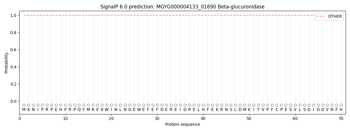
| Other | SP_Sec_SPI | LIPO_Sec_SPII | TAT_Tat_SPI | TATLIP_Sec_SPII | PILIN_Sec_SPIII |
|---|---|---|---|---|---|
| 1.000045 | 0.000000 | 0.000000 | 0.000000 | 0.000000 | 0.000000 |