You are browsing environment: HUMAN GUT
CAZyme Information: MGYG000004230_00038
You are here: Home > Sequence: MGYG000004230_00038
Basic Information |
Genomic context |
Full Sequence |
Enzyme annotations |
CAZy signature domains |
CDD domains |
CAZyme hits |
PDB hits |
Swiss-Prot hits |
SignalP and Lipop annotations |
TMHMM annotations
Basic Information help
| Species | Clostridium sp900759995 | |||||||||||
|---|---|---|---|---|---|---|---|---|---|---|---|---|
| Lineage | Bacteria; Firmicutes_A; Clostridia; Clostridiales; Clostridiaceae; Clostridium; Clostridium sp900759995 | |||||||||||
| CAZyme ID | MGYG000004230_00038 | |||||||||||
| CAZy Family | GH1 | |||||||||||
| CAZyme Description | Aryl-phospho-beta-D-glucosidase BglH | |||||||||||
| CAZyme Property |
|
|||||||||||
| Genome Property |
|
|||||||||||
| Gene Location | Start: 48252; End: 49691 Strand: + | |||||||||||
CAZyme Signature Domains help
| Family | Start | End | Evalue | family coverage |
|---|---|---|---|---|
| GH1 | 6 | 476 | 6.9e-153 | 0.9906759906759907 |
CDD Domains download full data without filtering help
| Cdd ID | Domain | E-Value | qStart | qEnd | sStart | sEnd | Domain Description |
|---|---|---|---|---|---|---|---|
| PRK09589 | celA | 0.0 | 7 | 479 | 3 | 476 | 6-phospho-beta-glucosidase; Reviewed |
| PRK09852 | PRK09852 | 0.0 | 6 | 479 | 2 | 473 | cryptic 6-phospho-beta-glucosidase; Provisional |
| PRK15014 | PRK15014 | 0.0 | 7 | 479 | 5 | 477 | 6-phospho-beta-glucosidase BglA; Provisional |
| COG2723 | BglB | 0.0 | 6 | 478 | 2 | 456 | Beta-glucosidase/6-phospho-beta-glucosidase/beta-galactosidase [Carbohydrate transport and metabolism]. |
| PRK09593 | arb | 0.0 | 8 | 479 | 6 | 477 | 6-phospho-beta-glucosidase; Reviewed |
CAZyme Hits help
| Hit ID | E-Value | Query Start | Query End | Hit Start | Hit End |
|---|---|---|---|---|---|
| QES73435.1 | 1.27e-309 | 8 | 479 | 5 | 476 |
| CDM68913.1 | 9.18e-305 | 8 | 479 | 5 | 476 |
| AOR24964.2 | 6.98e-284 | 8 | 479 | 7 | 478 |
| QKE74177.1 | 4.46e-264 | 1 | 479 | 1 | 479 |
| QDP39865.1 | 6.75e-261 | 7 | 479 | 5 | 477 |
PDB Hits download full data without filtering help
| Hit ID | E-Value | Query Start | Query End | Hit Start | Hit End | Description |
|---|---|---|---|---|---|---|
| 6WGD_A | 1.64e-232 | 1 | 479 | 1 | 469 | Crystalstructure of a 6-phospho-beta-glucosidase from Bacillus licheniformis [Bacillus licheniformis],6WGD_B Crystal structure of a 6-phospho-beta-glucosidase from Bacillus licheniformis [Bacillus licheniformis],6WGD_C Crystal structure of a 6-phospho-beta-glucosidase from Bacillus licheniformis [Bacillus licheniformis] |
| 4F66_A | 1.33e-193 | 6 | 479 | 5 | 480 | Thecrystal structure of 6-phospho-beta-glucosidase from Streptococcus mutans UA159 in complex with beta-D-glucose-6-phosphate. [Streptococcus mutans],4F66_B The crystal structure of 6-phospho-beta-glucosidase from Streptococcus mutans UA159 in complex with beta-D-glucose-6-phosphate. [Streptococcus mutans] |
| 4F79_A | 3.78e-193 | 6 | 479 | 5 | 480 | Thecrystal structure of 6-phospho-beta-glucosidase mutant (E375Q) in complex with Salicin 6-phosphate [Streptococcus mutans],4GPN_A The crystal structure of 6-P-beta-D-Glucosidase (E375Q mutant) from Streptococcus mutans UA150 in complex with Gentiobiose 6-phosphate. [Streptococcus mutans UA159],4GPN_B The crystal structure of 6-P-beta-D-Glucosidase (E375Q mutant) from Streptococcus mutans UA150 in complex with Gentiobiose 6-phosphate. [Streptococcus mutans UA159] |
| 2XHY_A | 2.42e-191 | 9 | 479 | 9 | 479 | CrystalStructure of E.coli BglA [Escherichia coli K-12],2XHY_B Crystal Structure of E.coli BglA [Escherichia coli K-12],2XHY_C Crystal Structure of E.coli BglA [Escherichia coli K-12],2XHY_D Crystal Structure of E.coli BglA [Escherichia coli K-12] |
| 3PN8_A | 6.70e-189 | 6 | 479 | 5 | 480 | Thecrystal structure of 6-phospho-beta-glucosidase from Streptococcus mutans UA159 [Streptococcus mutans],3PN8_B The crystal structure of 6-phospho-beta-glucosidase from Streptococcus mutans UA159 [Streptococcus mutans] |
Swiss-Prot Hits download full data without filtering help
| Hit ID | E-Value | Query Start | Query End | Hit Start | Hit End | Description |
|---|---|---|---|---|---|---|
| P24240 | 3.43e-235 | 8 | 479 | 4 | 473 | 6-phospho-beta-glucosidase AscB OS=Escherichia coli (strain K12) OX=83333 GN=ascB PE=3 SV=2 |
| P40740 | 1.40e-228 | 1 | 479 | 1 | 469 | Aryl-phospho-beta-D-glucosidase BglH OS=Bacillus subtilis (strain 168) OX=224308 GN=bglH PE=1 SV=2 |
| P42973 | 1.13e-196 | 9 | 479 | 5 | 479 | Aryl-phospho-beta-D-glucosidase BglA OS=Bacillus subtilis (strain 168) OX=224308 GN=bglA PE=1 SV=1 |
| Q46130 | 3.69e-196 | 8 | 479 | 7 | 471 | 6-phospho-beta-glucosidase OS=Clostridium longisporum OX=1523 GN=abgA PE=3 SV=1 |
| Q48409 | 1.93e-191 | 8 | 479 | 4 | 462 | Phospho-cellobiase OS=Klebsiella oxytoca OX=571 GN=casB PE=3 SV=1 |
SignalP and Lipop Annotations help
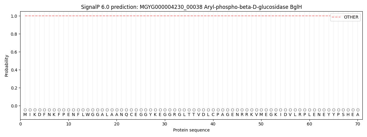
This protein is predicted as OTHER
| Other | SP_Sec_SPI | LIPO_Sec_SPII | TAT_Tat_SPI | TATLIP_Sec_SPII | PILIN_Sec_SPIII |
|---|---|---|---|---|---|
| 1.000061 | 0.000000 | 0.000000 | 0.000000 | 0.000000 | 0.000000 |