You are browsing environment: HUMAN GUT
CAZyme Information: MGYG000004275_01868
You are here: Home > Sequence: MGYG000004275_01868
Basic Information |
Genomic context |
Full Sequence |
Enzyme annotations |
CAZy signature domains |
CDD domains |
CAZyme hits |
PDB hits |
Swiss-Prot hits |
SignalP and Lipop annotations |
TMHMM annotations
Basic Information help
| Species | Ruminococcus_C sp000437175 | |||||||||||
|---|---|---|---|---|---|---|---|---|---|---|---|---|
| Lineage | Bacteria; Firmicutes_A; Clostridia; Oscillospirales; Ruminococcaceae; Ruminococcus_C; Ruminococcus_C sp000437175 | |||||||||||
| CAZyme ID | MGYG000004275_01868 | |||||||||||
| CAZy Family | GH26 | |||||||||||
| CAZyme Description | hypothetical protein | |||||||||||
| CAZyme Property |
|
|||||||||||
| Genome Property |
|
|||||||||||
| Gene Location | Start: 2074; End: 3666 Strand: - | |||||||||||
CAZyme Signature Domains help
| Family | Start | End | Evalue | family coverage |
|---|---|---|---|---|
| GH26 | 20 | 359 | 3.4e-98 | 0.9933993399339934 |
CDD Domains download full data without filtering help
| Cdd ID | Domain | E-Value | qStart | qEnd | sStart | sEnd | Domain Description |
|---|---|---|---|---|---|---|---|
| pfam02156 | Glyco_hydro_26 | 6.60e-45 | 20 | 359 | 2 | 311 | Glycosyl hydrolase family 26. |
| cd14256 | Dockerin_I | 7.29e-14 | 466 | 522 | 1 | 57 | Type I dockerin repeat domain. Bacterial cohesin domains bind to a complementary protein domain named dockerin, and this interaction is required for the formation of the cellulosome, a cellulose-degrading complex. The cellulosome consists of scaffoldin, a noncatalytic scaffolding polypeptide, that comprises repeating cohesion modules and a single carbohydrate-binding module (CBM). Specific calcium-dependent interactions between cohesins and dockerins appear to be essential for cellulosome assembly. This subfamily represents type I dockerins, which are responsible for anchoring a variety of enzymatic domains to the complex. |
| COG4124 | ManB2 | 3.34e-11 | 179 | 275 | 178 | 267 | Beta-mannanase [Carbohydrate transport and metabolism]. |
| cd14253 | Dockerin | 1.55e-07 | 467 | 522 | 1 | 56 | Dockerin repeat domain. Dockerins are modules in the cellulosome complex that often anchor catalytic subunits by binding to cohesin domains of scaffolding proteins. Three types of dockerins and their corresponding cohesin have been described in the literature. This alignment models two consecutive dockerin repeats, the functional unit. |
| pfam00404 | Dockerin_1 | 8.41e-07 | 467 | 521 | 1 | 55 | Dockerin type I repeat. The dockerin repeat is the binding partner of the cohesin domain pfam00963. The cohesin-dockerin interaction is the crucial interaction for complex formation in the cellulosome. The dockerin repeats, each bearing homology to the EF-hand calcium-binding loop bind calcium. |
CAZyme Hits help
| Hit ID | E-Value | Query Start | Query End | Hit Start | Hit End |
|---|---|---|---|---|---|
| ADU21037.1 | 3.37e-193 | 1 | 365 | 154 | 525 |
| QOS82751.1 | 4.01e-158 | 17 | 361 | 169 | 508 |
| APO46210.1 | 1.19e-157 | 17 | 361 | 169 | 508 |
| AZK49093.1 | 8.90e-157 | 17 | 361 | 169 | 508 |
| ASA20068.1 | 1.26e-156 | 17 | 370 | 169 | 517 |
PDB Hits download full data without filtering help
| Hit ID | E-Value | Query Start | Query End | Hit Start | Hit End | Description |
|---|---|---|---|---|---|---|
| 6D2X_A | 3.85e-54 | 18 | 360 | 4 | 334 | Crystalstructure of the GH26 domain from PbGH26-GH5A endo-beta-mannanase/endo-beta-glucanase from Prevotella bryantii [Prevotella bryantii B14] |
| 3ZM8_A | 5.30e-53 | 7 | 360 | 135 | 447 | ChainA, Gh26 Endo-beta-1,4-mannanase [Podospora anserina S mat+] |
| 6D2W_A | 8.03e-51 | 18 | 360 | 95 | 425 | Crystalstructure of Prevotella bryantii endo-beta-mannanase/endo-beta-glucanase PbGH26A-GH5A [Prevotella bryantii B14],6D2W_B Crystal structure of Prevotella bryantii endo-beta-mannanase/endo-beta-glucanase PbGH26A-GH5A [Prevotella bryantii B14] |
| 6HPF_A | 3.44e-42 | 22 | 360 | 10 | 310 | Structureof Inactive E165Q mutant of fungal non-CBM carrying GH26 endo-b-mannanase from Yunnania penicillata in complex with alpha-62-61-di-galactosyl-mannotriose [Yunnania penicillata] |
| 3WDQ_A | 1.72e-39 | 28 | 361 | 44 | 353 | Crystalstructure of beta-mannanase from a symbiotic protist of the termite Reticulitermes speratus [Symbiotic protist of Reticulitermes speratus],3WDR_A Crystal structure of beta-mannanase from a symbiotic protist of the termite Reticulitermes speratus complexed with gluco-manno-oligosaccharide [Symbiotic protist of Reticulitermes speratus] |
Swiss-Prot Hits download full data without filtering help
| Hit ID | E-Value | Query Start | Query End | Hit Start | Hit End | Description |
|---|---|---|---|---|---|---|
| P49425 | 1.08e-54 | 18 | 361 | 146 | 458 | Mannan endo-1,4-beta-mannosidase OS=Rhodothermus marinus (strain ATCC 43812 / DSM 4252 / R-10) OX=518766 GN=manA PE=1 SV=3 |
| G2Q4H7 | 1.24e-53 | 7 | 360 | 164 | 476 | Mannan endo-1,4-beta-mannosidase OS=Myceliophthora thermophila (strain ATCC 42464 / BCRC 31852 / DSM 1799) OX=573729 GN=Man26A PE=1 SV=1 |
| A2R6F5 | 2.40e-45 | 20 | 357 | 30 | 330 | Mannan endo-1,4-beta-mannosidase man26A OS=Aspergillus niger (strain CBS 513.88 / FGSC A1513) OX=425011 GN=man26A PE=1 SV=1 |
| P55298 | 2.00e-42 | 18 | 361 | 156 | 460 | Mannan endo-1,4-beta-mannosidase C OS=Piromyces sp. OX=45796 GN=MANC PE=2 SV=1 |
| Q5AWB7 | 2.76e-42 | 22 | 360 | 32 | 350 | Probable mannan endo-1,4-beta-mannosidase E OS=Emericella nidulans (strain FGSC A4 / ATCC 38163 / CBS 112.46 / NRRL 194 / M139) OX=227321 GN=manE PE=3 SV=1 |
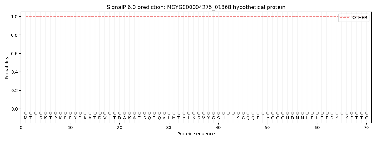
SignalP and Lipop Annotations help
This protein is predicted as OTHER
| Other | SP_Sec_SPI | LIPO_Sec_SPII | TAT_Tat_SPI | TATLIP_Sec_SPII | PILIN_Sec_SPIII |
|---|---|---|---|---|---|
| 1.000028 | 0.000002 | 0.000000 | 0.000000 | 0.000000 | 0.000000 |