You are browsing environment: HUMAN GUT
CAZyme Information: MGYG000004295_02326
You are here: Home > Sequence: MGYG000004295_02326
Basic Information |
Genomic context |
Full Sequence |
Enzyme annotations |
CAZy signature domains |
CDD domains |
CAZyme hits |
PDB hits |
Swiss-Prot hits |
SignalP and Lipop annotations |
TMHMM annotations
Basic Information help
| Species | Providencia rettgeri | |||||||||||
|---|---|---|---|---|---|---|---|---|---|---|---|---|
| Lineage | Bacteria; Proteobacteria; Gammaproteobacteria; Enterobacterales; Enterobacteriaceae; Providencia; Providencia rettgeri | |||||||||||
| CAZyme ID | MGYG000004295_02326 | |||||||||||
| CAZy Family | AA10 | |||||||||||
| CAZyme Description | GlcNAc-binding protein A | |||||||||||
| CAZyme Property |
|
|||||||||||
| Genome Property |
|
|||||||||||
| Gene Location | Start: 15793; End: 16371 Strand: - | |||||||||||
CAZyme Signature Domains help
| Family | Start | End | Evalue | family coverage |
|---|---|---|---|---|
| AA10 | 25 | 189 | 2e-51 | 0.9887640449438202 |
CDD Domains download full data without filtering help
| Cdd ID | Domain | E-Value | qStart | qEnd | sStart | sEnd | Domain Description |
|---|---|---|---|---|---|---|---|
| PRK13211 | PRK13211 | 4.97e-88 | 1 | 192 | 1 | 194 | N-acetylglucosamine-binding protein GbpA. |
| cd21177 | LPMO_AA10 | 5.87e-69 | 25 | 188 | 1 | 178 | lytic polysaccharide monooxygenase (LPMO) auxiliary activity family 10 (AA10). AA10 proteins are copper-dependent lytic polysaccharide monooxygenases (LPMOs), which may act on chitin or cellulose. The family used to be called CBM33. Activities in this family include lytic cellulose monooxygenase (C1-hydroxylating) (EC 1.14.99.54), lytic cellulose monooxygenase (C4-dehydrogenating) (EC 1.14.99.56), lytic chitin monooxygenase (EC 1.14.99.53), and lytic xylan monooxygenase/xylan oxidase (glycosidic bond-cleaving) (EC 1.14.99.-). Also included are viral chitin-binding glycoproteins such as fusolin and spheroidin-like proteins. |
| COG3397 | COG3397 | 1.06e-65 | 1 | 192 | 1 | 208 | Predicted carbohydrate-binding protein, contains CBM5 and CBM33 domains [General function prediction only]. |
| pfam03067 | LPMO_10 | 2.24e-53 | 25 | 188 | 1 | 185 | Lytic polysaccharide mono-oxygenase, cellulose-degrading. This domain is found associated with a wide variety of cellulose binding domains. This is a family of two very closely related proteins that together act as both a C1- and a C4-oxidising lytic polysaccharide mono-oxygenase, degrading cellulose. This domain is also found in baculoviral spheroidins and spindolins, protein of unknown function. |
CAZyme Hits help
| Hit ID | E-Value | Query Start | Query End | Hit Start | Hit End |
|---|---|---|---|---|---|
| AWS51697.1 | 2.38e-145 | 1 | 192 | 1 | 192 |
| QZY62658.1 | 2.38e-145 | 1 | 192 | 1 | 192 |
| QIF57910.1 | 1.38e-144 | 1 | 192 | 1 | 192 |
| QIF61950.1 | 1.38e-144 | 1 | 192 | 1 | 192 |
| BBU95908.1 | 1.63e-118 | 1 | 192 | 1 | 192 |
PDB Hits download full data without filtering help
| Hit ID | E-Value | Query Start | Query End | Hit Start | Hit End | Description |
|---|---|---|---|---|---|---|
| 2BEM_A | 6.24e-66 | 25 | 192 | 1 | 169 | Crystalstructure of the Serratia marcescens chitin-binding protein CBP21 [Serratia marcescens],2BEM_B Crystal structure of the Serratia marcescens chitin-binding protein CBP21 [Serratia marcescens],2BEM_C Crystal structure of the Serratia marcescens chitin-binding protein CBP21 [Serratia marcescens],2LHS_A Structure of the chitin binding protein 21 (CBP21) [Serratia marcescens] |
| 2BEN_A | 1.45e-64 | 25 | 192 | 1 | 169 | Crystalstructure of the Serratia marcescens chitin-binding protein CBP21 Y54A mutant. [Serratia marcescens],2BEN_B Crystal structure of the Serratia marcescens chitin-binding protein CBP21 Y54A mutant. [Serratia marcescens] |
| 6T5Z_A | 1.91e-63 | 25 | 192 | 1 | 172 | Crystalstructure of an AA10 LPMO from Photorhabdus luminescens [Photorhabdus laumondii subsp. laumondii TTO1],6T5Z_B Crystal structure of an AA10 LPMO from Photorhabdus luminescens [Photorhabdus laumondii subsp. laumondii TTO1],6T5Z_C Crystal structure of an AA10 LPMO from Photorhabdus luminescens [Photorhabdus laumondii subsp. laumondii TTO1] |
| 5WSZ_A | 3.78e-62 | 25 | 191 | 1 | 166 | Crystalstructure of a lytic polysaccharide monooxygenase,BtLPMO10A, from Bacillus thuringiensis [Bacillus thuringiensis serovar kurstaki],5WSZ_B Crystal structure of a lytic polysaccharide monooxygenase,BtLPMO10A, from Bacillus thuringiensis [Bacillus thuringiensis serovar kurstaki],5WSZ_C Crystal structure of a lytic polysaccharide monooxygenase,BtLPMO10A, from Bacillus thuringiensis [Bacillus thuringiensis serovar kurstaki],5WSZ_D Crystal structure of a lytic polysaccharide monooxygenase,BtLPMO10A, from Bacillus thuringiensis [Bacillus thuringiensis serovar kurstaki] |
| 2XWX_A | 5.60e-50 | 25 | 190 | 1 | 177 | Vibriocholerae colonization factor GbpA crystal structure [Vibrio cholerae],2XWX_B Vibrio cholerae colonization factor GbpA crystal structure [Vibrio cholerae] |
Swiss-Prot Hits download full data without filtering help
| Hit ID | E-Value | Query Start | Query End | Hit Start | Hit End | Description |
|---|---|---|---|---|---|---|
| Q8EHY2 | 5.24e-60 | 24 | 190 | 27 | 194 | GlcNAc-binding protein A OS=Shewanella oneidensis (strain MR-1) OX=211586 GN=gbpA PE=3 SV=2 |
| Q8D7V4 | 1.35e-49 | 10 | 190 | 9 | 199 | GlcNAc-binding protein A OS=Vibrio vulnificus (strain CMCP6) OX=216895 GN=gbpA PE=3 SV=1 |
| Q7MEW9 | 1.90e-49 | 10 | 190 | 9 | 199 | GlcNAc-binding protein A OS=Vibrio vulnificus (strain YJ016) OX=196600 GN=gbpA PE=3 SV=1 |
| Q87FT0 | 1.97e-49 | 1 | 190 | 1 | 200 | GlcNAc-binding protein A OS=Vibrio parahaemolyticus serotype O3:K6 (strain RIMD 2210633) OX=223926 GN=gbpA PE=3 SV=1 |
| A5F0H4 | 7.32e-49 | 21 | 190 | 20 | 200 | GlcNAc-binding protein A OS=Vibrio cholerae serotype O1 (strain ATCC 39541 / Classical Ogawa 395 / O395) OX=345073 GN=gbpA PE=3 SV=1 |
SignalP and Lipop Annotations help
This protein is predicted as SP
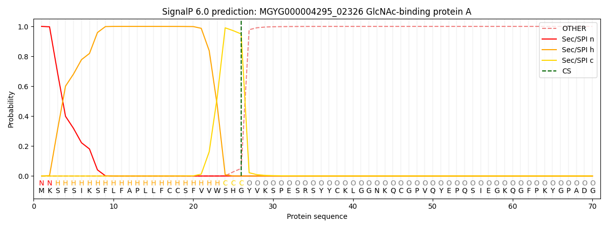
| Other | SP_Sec_SPI | LIPO_Sec_SPII | TAT_Tat_SPI | TATLIP_Sec_SPII | PILIN_Sec_SPIII |
|---|---|---|---|---|---|
| 0.000211 | 0.999251 | 0.000163 | 0.000135 | 0.000122 | 0.000118 |