You are browsing environment: HUMAN GUT
CAZyme Information: MGYG000004341_01319
You are here: Home > Sequence: MGYG000004341_01319
Basic Information |
Genomic context |
Full Sequence |
Enzyme annotations |
CAZy signature domains |
CDD domains |
CAZyme hits |
PDB hits |
Swiss-Prot hits |
SignalP and Lipop annotations |
TMHMM annotations
Basic Information help
| Species | CAG-110 sp000435995 | |||||||||||
|---|---|---|---|---|---|---|---|---|---|---|---|---|
| Lineage | Bacteria; Firmicutes_A; Clostridia; Oscillospirales; Oscillospiraceae; CAG-110; CAG-110 sp000435995 | |||||||||||
| CAZyme ID | MGYG000004341_01319 | |||||||||||
| CAZy Family | CBM50 | |||||||||||
| CAZyme Description | hypothetical protein | |||||||||||
| CAZyme Property |
|
|||||||||||
| Genome Property |
|
|||||||||||
| Gene Location | Start: 44719; End: 45678 Strand: + | |||||||||||
CDD Domains download full data without filtering help
| Cdd ID | Domain | E-Value | qStart | qEnd | sStart | sEnd | Domain Description |
|---|---|---|---|---|---|---|---|
| pfam01510 | Amidase_2 | 1.19e-24 | 23 | 150 | 1 | 121 | N-acetylmuramoyl-L-alanine amidase. This family includes zinc amidases that have N-acetylmuramoyl-L-alanine amidase activity EC:3.5.1.28. This enzyme domain cleaves the amide bond between N-acetylmuramoyl and L-amino acids in bacterial cell walls (preferentially: D-lactyl-L-Ala). The structure is known for the bacteriophage T7 structure and shows that two of the conserved histidines are zinc binding. |
| pfam08230 | CW_7 | 2.51e-18 | 279 | 318 | 1 | 40 | CW_7 repeat. This domain was originally found in the C-terminal moiety of the Cpl-7 lysozyme encoded by the Streptococcus pneumoniae bacteriophage Cp-7. It is also found in the cell wall hydrolases of human and life-stock pathogens. CW_7 repeats make up a cell wall binding motif. |
| cd00118 | LysM | 6.19e-17 | 229 | 272 | 2 | 45 | Lysin Motif is a small domain involved in binding peptidoglycan. LysM, a small globular domain with approximately 40 amino acids, is a widespread protein module involved in binding peptidoglycan in bacteria and chitin in eukaryotes. The domain was originally identified in enzymes that degrade bacterial cell walls, but proteins involved in many other biological functions also contain this domain. It has been reported that the LysM domain functions as a signal for specific plant-bacteria recognition in bacterial pathogenesis. Many of these enzymes are modular and are composed of catalytic units linked to one or several repeats of LysM domains. LysM domains are found in bacteria and eukaryotes. |
| pfam01476 | LysM | 1.04e-16 | 230 | 273 | 1 | 43 | LysM domain. The LysM (lysin motif) domain is about 40 residues long. It is found in a variety of enzymes involved in bacterial cell wall degradation. This domain may have a general peptidoglycan binding function. The structure of this domain is known. |
| cd06583 | PGRP | 4.71e-16 | 23 | 151 | 1 | 126 | Peptidoglycan recognition proteins (PGRPs) are pattern recognition receptors that bind, and in certain cases, hydrolyze peptidoglycans (PGNs) of bacterial cell walls. PGRPs have been divided into three classes: short PGRPs (PGRP-S), that are small (20 kDa) extracellular proteins; intermediate PGRPs (PGRP-I) that are 40-45 kDa and are predicted to be transmembrane proteins; and long PGRPs (PGRP-L), up to 90 kDa, which may be either intracellular or transmembrane. Several structures of PGRPs are known in insects and mammals, some bound with substrates like Muramyl Tripeptide (MTP) or Tracheal Cytotoxin (TCT). The substrate binding site is conserved in PGRP-LCx, PGRP-LE, and PGRP-Ialpha proteins. This family includes Zn-dependent N-Acetylmuramoyl-L-alanine Amidase, EC:3.5.1.28. This enzyme cleaves the amide bond between N-acetylmuramoyl and L-amino acids, preferentially D-lactyl-L-Ala, in bacterial cell walls. The structure for the bacteriophage T7 lysozyme shows that two of the conserved histidines and a cysteine are zinc binding residues. Site-directed mutagenesis of T7 lysozyme indicates that two conserved residues, a Tyr and a Lys, are important for amidase activity. |
CAZyme Hits help
| Hit ID | E-Value | Query Start | Query End | Hit Start | Hit End |
|---|---|---|---|---|---|
| ARE87697.1 | 8.64e-73 | 1 | 204 | 1 | 202 |
| AFB76124.1 | 2.27e-64 | 1 | 274 | 6 | 242 |
| QUO22979.1 | 1.23e-37 | 3 | 274 | 188 | 505 |
| ADB89215.1 | 7.87e-37 | 3 | 194 | 15 | 218 |
| AFB75734.1 | 8.66e-37 | 3 | 274 | 188 | 505 |
PDB Hits download full data without filtering help
| Hit ID | E-Value | Query Start | Query End | Hit Start | Hit End | Description |
|---|---|---|---|---|---|---|
| 4CVD_A | 1.14e-07 | 281 | 318 | 4 | 41 | ChainA, LYSOZYME [Streptococcus phage CP-7] |
| 5I8L_A | 5.25e-07 | 276 | 318 | 6 | 48 | ChainA, Lysozyme [Streptococcus phage CP-7] |
Swiss-Prot Hits download full data without filtering help
| Hit ID | E-Value | Query Start | Query End | Hit Start | Hit End | Description |
|---|---|---|---|---|---|---|
| Q49UX4 | 1.36e-07 | 228 | 277 | 86 | 134 | N-acetylmuramoyl-L-alanine amidase sle1 OS=Staphylococcus saprophyticus subsp. saprophyticus (strain ATCC 15305 / DSM 20229 / NCIMB 8711 / NCTC 7292 / S-41) OX=342451 GN=sle1 PE=3 SV=1 |
| Q5HRU2 | 1.03e-06 | 225 | 273 | 144 | 191 | N-acetylmuramoyl-L-alanine amidase sle1 OS=Staphylococcus epidermidis (strain ATCC 35984 / RP62A) OX=176279 GN=sle1 PE=3 SV=1 |
| Q8CMN2 | 1.03e-06 | 225 | 273 | 144 | 191 | N-acetylmuramoyl-L-alanine amidase sle1 OS=Staphylococcus epidermidis (strain ATCC 12228 / FDA PCI 1200) OX=176280 GN=sle1 PE=3 SV=1 |
| Q4L3C1 | 1.88e-06 | 223 | 277 | 83 | 136 | N-acetylmuramoyl-L-alanine amidase sle1 OS=Staphylococcus haemolyticus (strain JCSC1435) OX=279808 GN=sle1 PE=3 SV=1 |
| Q6GHI8 | 2.17e-06 | 226 | 279 | 173 | 226 | Probable cell wall hydrolase LytN OS=Staphylococcus aureus (strain MRSA252) OX=282458 GN=lytN PE=3 SV=2 |
SignalP and Lipop Annotations help
This protein is predicted as OTHER
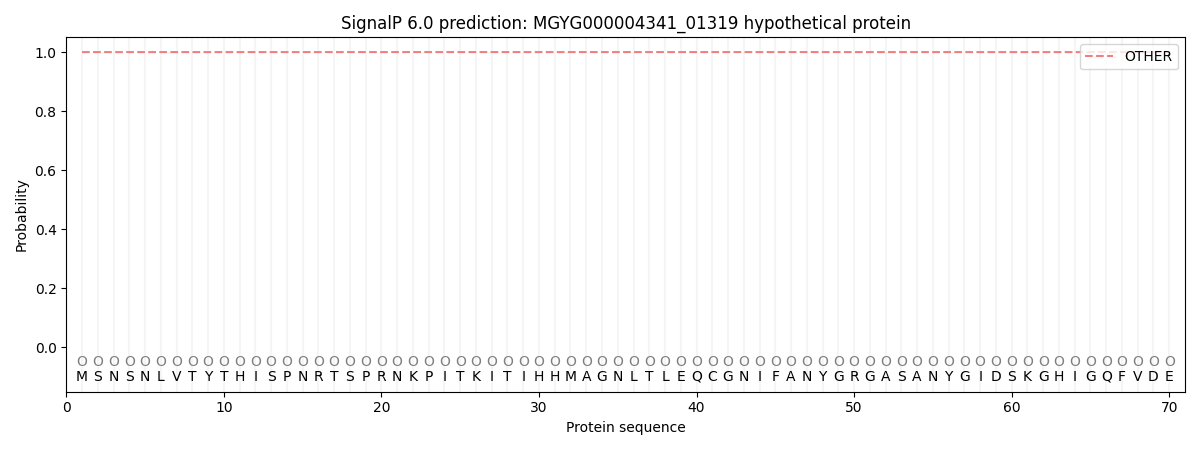
| Other | SP_Sec_SPI | LIPO_Sec_SPII | TAT_Tat_SPI | TATLIP_Sec_SPII | PILIN_Sec_SPIII |
|---|---|---|---|---|---|
| 1.000045 | 0.000001 | 0.000000 | 0.000000 | 0.000000 | 0.000000 |