You are browsing environment: HUMAN GUT
CAZyme Information: MGYG000004395_00381
You are here: Home > Sequence: MGYG000004395_00381
Basic Information |
Genomic context |
Full Sequence |
Enzyme annotations |
CAZy signature domains |
CDD domains |
CAZyme hits |
PDB hits |
Swiss-Prot hits |
SignalP and Lipop annotations |
TMHMM annotations
Basic Information help
| Species | ||||||||||||
|---|---|---|---|---|---|---|---|---|---|---|---|---|
| Lineage | Bacteria; Firmicutes; Bacilli; RF39; UBA660; CAG-594; | |||||||||||
| CAZyme ID | MGYG000004395_00381 | |||||||||||
| CAZy Family | GT0 | |||||||||||
| CAZyme Description | UDP-2,3-diacetamido-2,3-dideoxy-D-glucuronate 2-epimerase | |||||||||||
| CAZyme Property |
|
|||||||||||
| Genome Property |
|
|||||||||||
| Gene Location | Start: 48332; End: 49441 Strand: + | |||||||||||
CDD Domains download full data without filtering help
| Cdd ID | Domain | E-Value | qStart | qEnd | sStart | sEnd | Domain Description |
|---|---|---|---|---|---|---|---|
| cd03786 | GTB_UDP-GlcNAc_2-Epimerase | 1.65e-146 | 2 | 355 | 1 | 365 | UDP-N-acetylglucosamine 2-epimerase and similar proteins. Bacterial members of the UDP-N-Acetylglucosamine (GlcNAc) 2-Epimerase family (EC 5.1.3.14) are known to catalyze the reversible interconversion of UDP-GlcNAc and UDP-N-acetylmannosamine (UDP-ManNAc). The enzyme serves to produce an activated form of ManNAc residues (UDP-ManNAc) for use in the biosynthesis of a variety of cell surface polysaccharides; The mammalian enzyme is bifunctional, catalyzing both the inversion of stereochemistry at C-2 and the hydrolysis of the UDP-sugar linkage to generate free ManNAc. It also catalyzes the phosphorylation of ManNAc to generate ManNAc 6-phosphate, a precursor to salic acids. In mammals, sialic acids are found at the termini of oligosaccharides in a large variety of cell surface glycoconjugates and are key mediators of cell-cell recognition events. Mutations in human members of this family have been associated with Sialuria, a rare disease caused by the disorders of sialic acid metabolism. This family belongs to the GT-B structural superfamily of glycoslytransferases, which have characteristic N- and C-terminal domains each containing a typical Rossmann fold. The two domains have high structural homology despite minimal sequence homology. The large cleft that separates the two domains includes the catalytic center and permits a high degree of flexibility. |
| COG0381 | WecB | 4.65e-141 | 1 | 369 | 4 | 383 | UDP-N-acetylglucosamine 2-epimerase [Cell wall/membrane/envelope biogenesis]. |
| pfam02350 | Epimerase_2 | 5.59e-111 | 21 | 354 | 1 | 335 | UDP-N-acetylglucosamine 2-epimerase. This family consists of UDP-N-acetylglucosamine 2-epimerases EC:5.1.3.14 this enzyme catalyzes the production of UDP-ManNAc from UDP-GlcNAc. Note that some of the enzymes is this family are bifunctional, in these instances Pfam matches only the N-terminal half of the protein suggesting that the additional C-terminal part (when compared to mono-functional members of this family) is responsible for the UPD-N-acetylmannosamine kinase activity of these enzymes. This hypothesis is further supported by the assumption that the C-terminal part of rat Gne is the kinase domain. |
| TIGR00236 | wecB | 3.78e-75 | 1 | 357 | 1 | 364 | UDP-N-acetylglucosamine 2-epimerase. This cytosolic enzyme converts UDP-N-acetyl-D-glucosamine to UDP-N-acetyl-D-mannosamine. In E. coli, this is the first step in the pathway of enterobacterial common antigen biosynthesis.Members of this orthology group have many gene symbols, often reflecting the overall activity of the pathway and/or operon that includes it. Symbols include epsC (exopolysaccharide C) in Burkholderia solanacerum, cap8P (type 8 capsule P) in Staphylococcus aureus, and nfrC in an older designation based on the effects of deletion on phage N4 adsorption. Epimerase activity was also demonstrated in a bifunctional rat enzyme, for which the N-terminal domain appears to be orthologous. The set of proteins found above the suggested cutoff includes E. coli WecB in one of two deeply branched clusters and the rat UDP-N-acetylglucosamine 2-epimerase domain in the other. [Cell envelope, Biosynthesis and degradation of surface polysaccharides and lipopolysaccharides] |
| pfam03033 | Glyco_transf_28 | 2.52e-04 | 3 | 116 | 2 | 119 | Glycosyltransferase family 28 N-terminal domain. The glycosyltransferase family 28 includes monogalactosyldiacylglycerol synthase (EC 2.4.1.46) and UDP-N-acetylglucosamine transferase (EC 2.4.1.-). This N-terminal domain contains the acceptor binding site and likely membrane association site. This family also contains a large number of proteins that probably have quite distinct activities. |
CAZyme Hits help
| Hit ID | E-Value | Query Start | Query End | Hit Start | Hit End |
|---|---|---|---|---|---|
| AOP03843.1 | 6.84e-115 | 2 | 368 | 22 | 402 |
| AOP02687.1 | 6.84e-115 | 2 | 368 | 22 | 402 |
| AOP03732.1 | 6.84e-115 | 2 | 368 | 22 | 402 |
| AOP02662.1 | 6.84e-115 | 2 | 368 | 22 | 402 |
| AOP03614.1 | 6.84e-115 | 2 | 368 | 22 | 402 |
PDB Hits download full data without filtering help
| Hit ID | E-Value | Query Start | Query End | Hit Start | Hit End | Description |
|---|---|---|---|---|---|---|
| 4HWG_A | 3.10e-150 | 1 | 369 | 10 | 385 | Structureof UDP-N-acetylglucosamine 2-epimerase from Rickettsia bellii [Rickettsia bellii RML369-C] |
| 4NEQ_A | 1.47e-70 | 2 | 361 | 2 | 367 | Thestructure of UDP-GlcNAc 2-epimerase from Methanocaldococcus jannaschii [Methanocaldococcus jannaschii DSM 2661],4NES_A Crystal structure of Methanocaldococcus jannaschii UDP-GlcNAc 2-epimerase in complex with UDP-GlcNAc and UDP [Methanocaldococcus jannaschii DSM 2661] |
| 5ENZ_A | 9.14e-46 | 2 | 332 | 3 | 336 | S.aureus MnaA-UDP co-structure [Staphylococcus aureus],5ENZ_B S. aureus MnaA-UDP co-structure [Staphylococcus aureus] |
| 4FKZ_A | 3.18e-39 | 1 | 321 | 4 | 326 | Crystalstructure of Bacillus subtilis UDP-GlcNAc 2-epimerase in complex with UDP-GlcNAc and UDP [Bacillus subtilis subsp. subtilis str. 168],4FKZ_B Crystal structure of Bacillus subtilis UDP-GlcNAc 2-epimerase in complex with UDP-GlcNAc and UDP [Bacillus subtilis subsp. subtilis str. 168] |
| 3BEO_A | 1.31e-37 | 1 | 332 | 9 | 343 | AStructural Basis for the allosteric regulation of non-hydrolyzing UDP-GlcNAc 2-epimerases [Bacillus anthracis],3BEO_B A Structural Basis for the allosteric regulation of non-hydrolyzing UDP-GlcNAc 2-epimerases [Bacillus anthracis] |
Swiss-Prot Hits download full data without filtering help
| Hit ID | E-Value | Query Start | Query End | Hit Start | Hit End | Description |
|---|---|---|---|---|---|---|
| Q58899 | 1.03e-71 | 1 | 354 | 1 | 361 | UDP-N-acetylglucosamine 2-epimerase OS=Methanocaldococcus jannaschii (strain ATCC 43067 / DSM 2661 / JAL-1 / JCM 10045 / NBRC 100440) OX=243232 GN=wecB PE=1 SV=1 |
| Q6LZC4 | 6.41e-70 | 2 | 330 | 3 | 338 | UDP-N-acetylglucosamine 2-epimerase OS=Methanococcus maripaludis (strain S2 / LL) OX=267377 GN=wecB PE=1 SV=1 |
| Q6M0B4 | 6.77e-67 | 1 | 325 | 1 | 327 | UDP-N-acetylglucosamine 2-epimerase homolog OS=Methanococcus maripaludis (strain S2 / LL) OX=267377 GN=MMP0357 PE=1 SV=1 |
| G3XD61 | 3.56e-50 | 1 | 324 | 1 | 322 | UDP-2,3-diacetamido-2,3-dideoxy-D-glucuronate 2-epimerase OS=Pseudomonas aeruginosa (strain ATCC 15692 / DSM 22644 / CIP 104116 / JCM 14847 / LMG 12228 / 1C / PRS 101 / PAO1) OX=208964 GN=wbpI PE=1 SV=1 |
| P39131 | 1.49e-38 | 1 | 321 | 4 | 326 | UDP-N-acetylglucosamine 2-epimerase OS=Bacillus subtilis (strain 168) OX=224308 GN=mnaA PE=1 SV=1 |
SignalP and Lipop Annotations help
This protein is predicted as OTHER
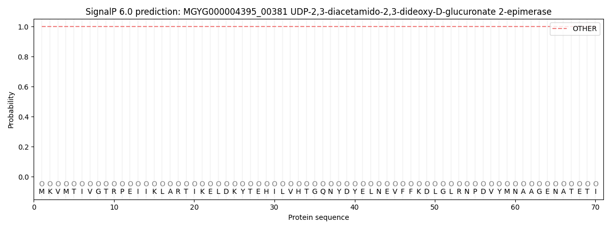
| Other | SP_Sec_SPI | LIPO_Sec_SPII | TAT_Tat_SPI | TATLIP_Sec_SPII | PILIN_Sec_SPIII |
|---|---|---|---|---|---|
| 1.000048 | 0.000000 | 0.000000 | 0.000000 | 0.000000 | 0.000000 |