You are browsing environment: HUMAN GUT
CAZyme Information: MGYG000004435_00421
You are here: Home > Sequence: MGYG000004435_00421
Basic Information |
Genomic context |
Full Sequence |
Enzyme annotations |
CAZy signature domains |
CDD domains |
CAZyme hits |
PDB hits |
Swiss-Prot hits |
SignalP and Lipop annotations |
TMHMM annotations
Basic Information help
| Species | UBA7597 sp900767195 | |||||||||||
|---|---|---|---|---|---|---|---|---|---|---|---|---|
| Lineage | Bacteria; Firmicutes_A; Clostridia_A; Christensenellales; Borkfalkiaceae; UBA7597; UBA7597 sp900767195 | |||||||||||
| CAZyme ID | MGYG000004435_00421 | |||||||||||
| CAZy Family | GH109 | |||||||||||
| CAZyme Description | Alpha-N-acetylgalactosaminidase | |||||||||||
| CAZyme Property |
|
|||||||||||
| Genome Property |
|
|||||||||||
| Gene Location | Start: 36176; End: 37474 Strand: - | |||||||||||
CAZyme Signature Domains help
| Family | Start | End | Evalue | family coverage |
|---|---|---|---|---|
| GH109 | 1 | 398 | 6.9e-137 | 0.9874686716791979 |
CDD Domains download full data without filtering help
| Cdd ID | Domain | E-Value | qStart | qEnd | sStart | sEnd | Domain Description |
|---|---|---|---|---|---|---|---|
| COG0673 | MviM | 4.66e-20 | 1 | 160 | 1 | 157 | Predicted dehydrogenase [General function prediction only]. |
| pfam01408 | GFO_IDH_MocA | 9.57e-11 | 4 | 120 | 1 | 112 | Oxidoreductase family, NAD-binding Rossmann fold. This family of enzymes utilize NADP or NAD. This family is called the GFO/IDH/MOCA family in swiss-prot. |
| PRK08618 | PRK08618 | 2.95e-05 | 2 | 105 | 125 | 223 | ornithine cyclodeaminase family protein. |
| PRK10206 | PRK10206 | 0.001 | 61 | 152 | 55 | 146 | putative oxidoreductase; Provisional |
| cd05284 | arabinose_DH_like | 0.005 | 6 | 108 | 174 | 274 | D-arabinose dehydrogenase. This group contains arabinose dehydrogenase (AraDH) and related alcohol dehydrogenases. AraDH is a member of the medium chain dehydrogenase/reductase family and catalyzes the NAD(P)-dependent oxidation of D-arabinose and other pentoses, the initial step in the metabolism of d-arabinose into 2-oxoglutarate. Like the alcohol dehydrogenases, AraDH binds a zinc in the catalytic cleft as well as a distal structural zinc. AraDH forms homotetramers as a dimer of dimers. AraDH replaces a conserved catalytic His with replace with Arg, compared to the canonical ADH site. NAD(P)(H)-dependent oxidoreductases are the major enzymes in the interconversion of alcohols and aldehydes, or ketones. Alcohol dehydrogenase in the liver converts ethanol and NAD+ to acetaldehyde and NADH, while in yeast and some other microorganisms ADH catalyzes the conversion acetaldehyde to ethanol in alcoholic fermentation. ADH is a member of the medium chain alcohol dehydrogenase family (MDR), which has a NAD(P)(H)-binding domain in a Rossmann fold of a beta-alpha form. The NAD(H)-binding region is comprised of 2 structurally similar halves, each of which contacts a mononucleotide. A GxGxxG motif after the first mononucleotide contact half allows the close contact of the coenzyme with the ADH backbone. The N-terminal catalytic domain has a distant homology to GroES. These proteins typically form dimers (typically higher plants, mammals) or tetramers (yeast, bacteria), and have 2 tightly bound zinc atoms per subunit, a catalytic zinc at the active site and a structural zinc in a lobe of the catalytic domain. NAD(H) binding occurs in the cleft between the catalytic and coenzyme-binding domains at the active site, and coenzyme binding induces a conformational closing of this cleft. Coenzyme binding typically precedes and contributes to substrate binding. In human ADH catalysis, the zinc ion helps coordinate the alcohol, followed by deprotonation of a histidine, the ribose of NAD, a serine, then the alcohol, which allows the transfer of a hydride to NAD+, creating NADH and a zinc-bound aldehyde or ketone. In yeast and some bacteria, the active site zinc binds an aldehyde, polarizing it, and leading to the reverse reaction. |
CAZyme Hits help
| Hit ID | E-Value | Query Start | Query End | Hit Start | Hit End |
|---|---|---|---|---|---|
| ALS29040.1 | 2.42e-139 | 4 | 400 | 5 | 398 |
| QTH43530.1 | 3.94e-137 | 3 | 400 | 4 | 398 |
| QUO31207.1 | 1.01e-134 | 1 | 400 | 1 | 398 |
| QTH41319.1 | 2.93e-133 | 3 | 398 | 7 | 398 |
| AVM42018.1 | 8.04e-133 | 1 | 399 | 1 | 398 |
PDB Hits download full data without filtering help
| Hit ID | E-Value | Query Start | Query End | Hit Start | Hit End | Description |
|---|---|---|---|---|---|---|
| 2IXA_A | 1.68e-80 | 2 | 397 | 19 | 431 | A-zyme,N-acetylgalactosaminidase [Elizabethkingia meningoseptica],2IXB_A Crystal structure of N-ACETYLGALACTOSAMINIDASE in complex with GalNAC [Elizabethkingia meningoseptica] |
| 6T2B_A | 2.37e-63 | 3 | 408 | 42 | 449 | Glycosidehydrolase family 109 from Akkermansia muciniphila in complex with GalNAc and NAD+. [Akkermansia muciniphila],6T2B_B Glycoside hydrolase family 109 from Akkermansia muciniphila in complex with GalNAc and NAD+. [Akkermansia muciniphila],6T2B_C Glycoside hydrolase family 109 from Akkermansia muciniphila in complex with GalNAc and NAD+. [Akkermansia muciniphila],6T2B_D Glycoside hydrolase family 109 from Akkermansia muciniphila in complex with GalNAc and NAD+. [Akkermansia muciniphila] |
| 3EC7_A | 1.17e-13 | 4 | 158 | 24 | 176 | CrystalStructure of Putative Dehydrogenase from Salmonella typhimurium LT2 [Salmonella enterica subsp. enterica serovar Typhimurium str. LT2],3EC7_B Crystal Structure of Putative Dehydrogenase from Salmonella typhimurium LT2 [Salmonella enterica subsp. enterica serovar Typhimurium str. LT2],3EC7_C Crystal Structure of Putative Dehydrogenase from Salmonella typhimurium LT2 [Salmonella enterica subsp. enterica serovar Typhimurium str. LT2],3EC7_D Crystal Structure of Putative Dehydrogenase from Salmonella typhimurium LT2 [Salmonella enterica subsp. enterica serovar Typhimurium str. LT2],3EC7_E Crystal Structure of Putative Dehydrogenase from Salmonella typhimurium LT2 [Salmonella enterica subsp. enterica serovar Typhimurium str. LT2],3EC7_F Crystal Structure of Putative Dehydrogenase from Salmonella typhimurium LT2 [Salmonella enterica subsp. enterica serovar Typhimurium str. LT2],3EC7_G Crystal Structure of Putative Dehydrogenase from Salmonella typhimurium LT2 [Salmonella enterica subsp. enterica serovar Typhimurium str. LT2],3EC7_H Crystal Structure of Putative Dehydrogenase from Salmonella typhimurium LT2 [Salmonella enterica subsp. enterica serovar Typhimurium str. LT2] |
| 3EZY_A | 6.60e-10 | 4 | 150 | 3 | 144 | Crystalstructure of probable dehydrogenase TM_0414 from Thermotoga maritima [Thermotoga maritima],3EZY_B Crystal structure of probable dehydrogenase TM_0414 from Thermotoga maritima [Thermotoga maritima],3EZY_C Crystal structure of probable dehydrogenase TM_0414 from Thermotoga maritima [Thermotoga maritima],3EZY_D Crystal structure of probable dehydrogenase TM_0414 from Thermotoga maritima [Thermotoga maritima] |
| 3E18_A | 2.24e-09 | 1 | 157 | 3 | 156 | CRYSTALSTRUCTURE OF NAD-BINDING PROTEIN FROM Listeria innocua [Listeria innocua],3E18_B CRYSTAL STRUCTURE OF NAD-BINDING PROTEIN FROM Listeria innocua [Listeria innocua] |
Swiss-Prot Hits download full data without filtering help
| Hit ID | E-Value | Query Start | Query End | Hit Start | Hit End | Description |
|---|---|---|---|---|---|---|
| A4Q8F7 | 9.17e-80 | 2 | 397 | 19 | 431 | Alpha-N-acetylgalactosaminidase OS=Elizabethkingia meningoseptica OX=238 GN=nagA PE=1 SV=1 |
| B2FLK4 | 5.22e-78 | 2 | 400 | 32 | 444 | Glycosyl hydrolase family 109 protein OS=Stenotrophomonas maltophilia (strain K279a) OX=522373 GN=Smlt4431 PE=3 SV=1 |
| A4Q8G1 | 2.32e-76 | 4 | 400 | 53 | 462 | Alpha-N-acetylgalactosaminidase OS=Tannerella forsythia OX=28112 GN=nagA PE=3 SV=1 |
| A6KY05 | 5.37e-73 | 4 | 400 | 17 | 413 | Glycosyl hydrolase family 109 protein 2 OS=Phocaeicola vulgatus (strain ATCC 8482 / DSM 1447 / JCM 5826 / CCUG 4940 / NBRC 14291 / NCTC 11154) OX=435590 GN=BVU_0611 PE=3 SV=1 |
| Q01S58 | 2.94e-72 | 4 | 400 | 43 | 435 | Glycosyl hydrolase family 109 protein OS=Solibacter usitatus (strain Ellin6076) OX=234267 GN=Acid_6590 PE=3 SV=1 |
SignalP and Lipop Annotations help
This protein is predicted as OTHER
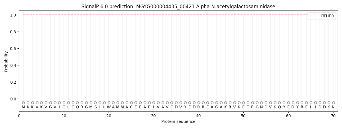
| Other | SP_Sec_SPI | LIPO_Sec_SPII | TAT_Tat_SPI | TATLIP_Sec_SPII | PILIN_Sec_SPIII |
|---|---|---|---|---|---|
| 1.000062 | 0.000000 | 0.000000 | 0.000000 | 0.000000 | 0.000000 |