You are browsing environment: HUMAN GUT
CAZyme Information: MGYG000004443_01504
You are here: Home > Sequence: MGYG000004443_01504
Basic Information |
Genomic context |
Full Sequence |
Enzyme annotations |
CAZy signature domains |
CDD domains |
CAZyme hits |
PDB hits |
Swiss-Prot hits |
SignalP and Lipop annotations |
TMHMM annotations
Basic Information help
| Species | ||||||||||||
|---|---|---|---|---|---|---|---|---|---|---|---|---|
| Lineage | Bacteria; Bacteroidota; Bacteroidia; Bacteroidales; Coprobacteraceae; ; | |||||||||||
| CAZyme ID | MGYG000004443_01504 | |||||||||||
| CAZy Family | GH109 | |||||||||||
| CAZyme Description | Inositol 2-dehydrogenase/D-chiro-inositol 3-dehydrogenase | |||||||||||
| CAZyme Property |
|
|||||||||||
| Genome Property |
|
|||||||||||
| Gene Location | Start: 6174; End: 7532 Strand: - | |||||||||||
CAZyme Signature Domains help
| Family | Start | End | Evalue | family coverage |
|---|---|---|---|---|
| GH109 | 65 | 252 | 3e-22 | 0.45614035087719296 |
CDD Domains download full data without filtering help
| Cdd ID | Domain | E-Value | qStart | qEnd | sStart | sEnd | Domain Description |
|---|---|---|---|---|---|---|---|
| COG0673 | MviM | 2.77e-36 | 64 | 416 | 3 | 338 | Predicted dehydrogenase [General function prediction only]. |
| pfam01408 | GFO_IDH_MocA | 2.65e-11 | 65 | 187 | 1 | 112 | Oxidoreductase family, NAD-binding Rossmann fold. This family of enzymes utilize NADP or NAD. This family is called the GFO/IDH/MOCA family in swiss-prot. |
| TIGR04380 | myo_inos_iolG | 4.10e-10 | 64 | 291 | 1 | 198 | inositol 2-dehydrogenase. All members of the seed alignment for this model are known or predicted inositol 2-dehydrogenase sequences co-clustered with other enzymes for catabolism of myo-inositol or closely related compounds. Inositol 2-dehydrogenase catalyzes the first step in inositol catabolism. Members of this family may vary somewhat in their ranges of acceptable substrates and some may act on analogs to myo-inositol rather than myo-inositol per se. [Energy metabolism, Sugars] |
| PRK10206 | PRK10206 | 9.81e-06 | 135 | 221 | 62 | 145 | putative oxidoreductase; Provisional |
| cd19930 | REC_DesR-like | 0.005 | 77 | 175 | 11 | 112 | phosphoacceptor receiver (REC) domain of DesR and similar proteins. This group is composed of Bacillus subtilis DesR, Streptococcus pneumoniae response regulator spr1814, and similar proteins, all containing an N-terminal REC domain and a C-terminal LuxR family helix-turn-helix (HTH) DNA-binding output domain. DesR is a response regulator that, together with its cognate sensor kinase DesK, comprises a two-component regulatory system that controls membrane fluidity. Phosphorylation of the REC domain of DesR is allosterically coupled to two distinct exposed surfaces of the protein, controlling noncanonical dimerization/tetramerization, cooperative activation, and DesK binding. REC domains function as phosphorylation-mediated switches within response regulators, but some also transfer phosphoryl groups in multistep phosphorelays. |
CAZyme Hits help
| Hit ID | E-Value | Query Start | Query End | Hit Start | Hit End |
|---|---|---|---|---|---|
| QGA23801.1 | 1.33e-240 | 9 | 452 | 5 | 453 |
| ABR45528.1 | 1.97e-225 | 6 | 449 | 2 | 454 |
| QRO18095.1 | 1.97e-225 | 6 | 449 | 2 | 454 |
| QUT52998.1 | 3.97e-225 | 6 | 449 | 2 | 454 |
| SCM55443.1 | 5.05e-225 | 6 | 452 | 2 | 456 |
PDB Hits download full data without filtering help
| Hit ID | E-Value | Query Start | Query End | Hit Start | Hit End | Description |
|---|---|---|---|---|---|---|
| 4N54_A | 7.43e-09 | 65 | 289 | 15 | 215 | ChainA, Inositol dehydrogenase [Lacticaseibacillus casei BL23],4N54_B Chain B, Inositol dehydrogenase [Lacticaseibacillus casei BL23],4N54_C Chain C, Inositol dehydrogenase [Lacticaseibacillus casei BL23],4N54_D Chain D, Inositol dehydrogenase [Lacticaseibacillus casei BL23] |
| 4MKX_A | 7.52e-09 | 65 | 289 | 18 | 218 | ChainA, Inositol dehydrogenase [Lacticaseibacillus casei BL23],4MKZ_A Chain A, Inositol dehydrogenase [Lacticaseibacillus casei BL23] |
| 3EC7_A | 3.17e-08 | 61 | 315 | 20 | 243 | CrystalStructure of Putative Dehydrogenase from Salmonella typhimurium LT2 [Salmonella enterica subsp. enterica serovar Typhimurium str. LT2],3EC7_B Crystal Structure of Putative Dehydrogenase from Salmonella typhimurium LT2 [Salmonella enterica subsp. enterica serovar Typhimurium str. LT2],3EC7_C Crystal Structure of Putative Dehydrogenase from Salmonella typhimurium LT2 [Salmonella enterica subsp. enterica serovar Typhimurium str. LT2],3EC7_D Crystal Structure of Putative Dehydrogenase from Salmonella typhimurium LT2 [Salmonella enterica subsp. enterica serovar Typhimurium str. LT2],3EC7_E Crystal Structure of Putative Dehydrogenase from Salmonella typhimurium LT2 [Salmonella enterica subsp. enterica serovar Typhimurium str. LT2],3EC7_F Crystal Structure of Putative Dehydrogenase from Salmonella typhimurium LT2 [Salmonella enterica subsp. enterica serovar Typhimurium str. LT2],3EC7_G Crystal Structure of Putative Dehydrogenase from Salmonella typhimurium LT2 [Salmonella enterica subsp. enterica serovar Typhimurium str. LT2],3EC7_H Crystal Structure of Putative Dehydrogenase from Salmonella typhimurium LT2 [Salmonella enterica subsp. enterica serovar Typhimurium str. LT2] |
| 5A02_A | 2.18e-07 | 103 | 344 | 38 | 262 | Crystalstructure of aldose-aldose oxidoreductase from Caulobacter crescentus complexed with glycerol [Caulobacter vibrioides CB15],5A02_B Crystal structure of aldose-aldose oxidoreductase from Caulobacter crescentus complexed with glycerol [Caulobacter vibrioides CB15],5A02_C Crystal structure of aldose-aldose oxidoreductase from Caulobacter crescentus complexed with glycerol [Caulobacter vibrioides CB15],5A02_D Crystal structure of aldose-aldose oxidoreductase from Caulobacter crescentus complexed with glycerol [Caulobacter vibrioides CB15],5A02_E Crystal structure of aldose-aldose oxidoreductase from Caulobacter crescentus complexed with glycerol [Caulobacter vibrioides CB15],5A02_F Crystal structure of aldose-aldose oxidoreductase from Caulobacter crescentus complexed with glycerol [Caulobacter vibrioides CB15],5A03_A Crystal structure of aldose-aldose oxidoreductase from Caulobacter crescentus complexed with xylose [Caulobacter vibrioides CB15],5A03_B Crystal structure of aldose-aldose oxidoreductase from Caulobacter crescentus complexed with xylose [Caulobacter vibrioides CB15],5A03_C Crystal structure of aldose-aldose oxidoreductase from Caulobacter crescentus complexed with xylose [Caulobacter vibrioides CB15],5A03_D Crystal structure of aldose-aldose oxidoreductase from Caulobacter crescentus complexed with xylose [Caulobacter vibrioides CB15],5A03_E Crystal structure of aldose-aldose oxidoreductase from Caulobacter crescentus complexed with xylose [Caulobacter vibrioides CB15],5A03_F Crystal structure of aldose-aldose oxidoreductase from Caulobacter crescentus complexed with xylose [Caulobacter vibrioides CB15],5A04_A Crystal structure of aldose-aldose oxidoreductase from Caulobacter crescentus complexed with glucose [Caulobacter vibrioides CB15],5A04_B Crystal structure of aldose-aldose oxidoreductase from Caulobacter crescentus complexed with glucose [Caulobacter vibrioides CB15],5A04_C Crystal structure of aldose-aldose oxidoreductase from Caulobacter crescentus complexed with glucose [Caulobacter vibrioides CB15],5A04_D Crystal structure of aldose-aldose oxidoreductase from Caulobacter crescentus complexed with glucose [Caulobacter vibrioides CB15],5A04_E Crystal structure of aldose-aldose oxidoreductase from Caulobacter crescentus complexed with glucose [Caulobacter vibrioides CB15],5A04_F Crystal structure of aldose-aldose oxidoreductase from Caulobacter crescentus complexed with glucose [Caulobacter vibrioides CB15],5A05_A Crystal structure of aldose-aldose oxidoreductase from Caulobacter crescentus complexed with maltotriose [Caulobacter vibrioides CB15],5A05_B Crystal structure of aldose-aldose oxidoreductase from Caulobacter crescentus complexed with maltotriose [Caulobacter vibrioides CB15],5A05_C Crystal structure of aldose-aldose oxidoreductase from Caulobacter crescentus complexed with maltotriose [Caulobacter vibrioides CB15],5A05_D Crystal structure of aldose-aldose oxidoreductase from Caulobacter crescentus complexed with maltotriose [Caulobacter vibrioides CB15],5A05_E Crystal structure of aldose-aldose oxidoreductase from Caulobacter crescentus complexed with maltotriose [Caulobacter vibrioides CB15],5A05_F Crystal structure of aldose-aldose oxidoreductase from Caulobacter crescentus complexed with maltotriose [Caulobacter vibrioides CB15],5A06_A Crystal structure of aldose-aldose oxidoreductase from Caulobacter crescentus complexed with sorbitol [Caulobacter vibrioides CB15],5A06_B Crystal structure of aldose-aldose oxidoreductase from Caulobacter crescentus complexed with sorbitol [Caulobacter vibrioides CB15],5A06_C Crystal structure of aldose-aldose oxidoreductase from Caulobacter crescentus complexed with sorbitol [Caulobacter vibrioides CB15],5A06_D Crystal structure of aldose-aldose oxidoreductase from Caulobacter crescentus complexed with sorbitol [Caulobacter vibrioides CB15],5A06_E Crystal structure of aldose-aldose oxidoreductase from Caulobacter crescentus complexed with sorbitol [Caulobacter vibrioides CB15],5A06_F Crystal structure of aldose-aldose oxidoreductase from Caulobacter crescentus complexed with sorbitol [Caulobacter vibrioides CB15] |
| 3E18_A | 2.28e-06 | 69 | 355 | 10 | 260 | CRYSTALSTRUCTURE OF NAD-BINDING PROTEIN FROM Listeria innocua [Listeria innocua],3E18_B CRYSTAL STRUCTURE OF NAD-BINDING PROTEIN FROM Listeria innocua [Listeria innocua] |
Swiss-Prot Hits download full data without filtering help
| Hit ID | E-Value | Query Start | Query End | Hit Start | Hit End | Description |
|---|---|---|---|---|---|---|
| Q01S58 | 1.43e-12 | 12 | 347 | 5 | 335 | Glycosyl hydrolase family 109 protein OS=Solibacter usitatus (strain Ellin6076) OX=234267 GN=Acid_6590 PE=3 SV=1 |
| P0C863 | 3.56e-11 | 42 | 234 | 33 | 220 | Glycosyl hydrolase family 109 protein 1 OS=Bacteroides fragilis (strain YCH46) OX=295405 GN=BF0931 PE=3 SV=1 |
| Q5LGZ0 | 3.56e-11 | 42 | 234 | 33 | 220 | Glycosyl hydrolase family 109 protein 1 OS=Bacteroides fragilis (strain ATCC 25285 / DSM 2151 / CCUG 4856 / JCM 11019 / NCTC 9343 / Onslow) OX=272559 GN=BF0853 PE=1 SV=1 |
| Q54728 | 1.04e-09 | 65 | 309 | 2 | 219 | Uncharacterized oxidoreductase SP_1686 OS=Streptococcus pneumoniae serotype 4 (strain ATCC BAA-334 / TIGR4) OX=170187 GN=SP_1686 PE=3 SV=2 |
| A8H2K3 | 1.05e-09 | 12 | 234 | 4 | 217 | Glycosyl hydrolase family 109 protein OS=Shewanella pealeana (strain ATCC 700345 / ANG-SQ1) OX=398579 GN=Spea_1465 PE=3 SV=1 |
SignalP and Lipop Annotations help
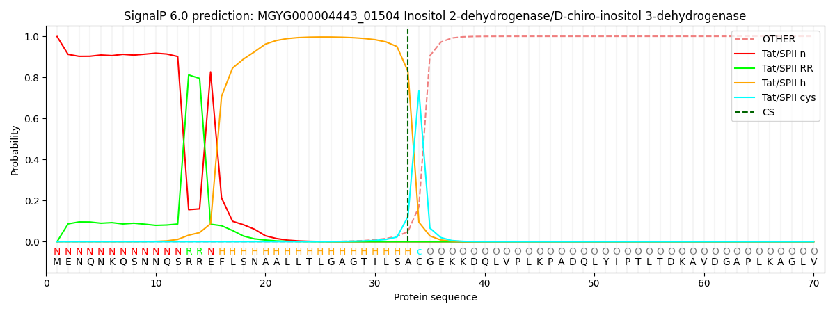
This protein is predicted as TATLIPO

| Other | SP_Sec_SPI | LIPO_Sec_SPII | TAT_Tat_SPI | TATLIP_Sec_SPII | PILIN_Sec_SPIII |
|---|---|---|---|---|---|
| 0.000000 | 0.000000 | 0.000039 | 0.001470 | 0.998490 | 0.000000 |
