You are browsing environment: HUMAN GUT
CAZyme Information: MGYG000004754_01668
You are here: Home > Sequence: MGYG000004754_01668
Basic Information |
Genomic context |
Full Sequence |
Enzyme annotations |
CAZy signature domains |
CDD domains |
CAZyme hits |
PDB hits |
Swiss-Prot hits |
SignalP and Lipop annotations |
TMHMM annotations
Basic Information help
| Species | Elizabethkingia bruuniana | |||||||||||
|---|---|---|---|---|---|---|---|---|---|---|---|---|
| Lineage | Bacteria; Bacteroidota; Bacteroidia; Flavobacteriales; Weeksellaceae; Elizabethkingia; Elizabethkingia bruuniana | |||||||||||
| CAZyme ID | MGYG000004754_01668 | |||||||||||
| CAZy Family | GT4 | |||||||||||
| CAZyme Description | hypothetical protein | |||||||||||
| CAZyme Property |
|
|||||||||||
| Genome Property |
|
|||||||||||
| Gene Location | Start: 732020; End: 733297 Strand: + | |||||||||||
CDD Domains download full data without filtering help
| Cdd ID | Domain | E-Value | qStart | qEnd | sStart | sEnd | Domain Description |
|---|---|---|---|---|---|---|---|
| cd03801 | GT4_PimA-like | 1.09e-19 | 111 | 358 | 134 | 357 | phosphatidyl-myo-inositol mannosyltransferase. This family is most closely related to the GT4 family of glycosyltransferases and named after PimA in Propionibacterium freudenreichii, which is involved in the biosynthesis of phosphatidyl-myo-inositol mannosides (PIM) which are early precursors in the biosynthesis of lipomannans (LM) and lipoarabinomannans (LAM), and catalyzes the addition of a mannosyl residue from GDP-D-mannose (GDP-Man) to the position 2 of the carrier lipid phosphatidyl-myo-inositol (PI) to generate a phosphatidyl-myo-inositol bearing an alpha-1,2-linked mannose residue (PIM1). Glycosyltransferases catalyze the transfer of sugar moieties from activated donor molecules to specific acceptor molecules, forming glycosidic bonds. The acceptor molecule can be a lipid, a protein, a heterocyclic compound, or another carbohydrate residue. This group of glycosyltransferases is most closely related to the previously defined glycosyltransferase family 1 (GT1). The members of this family may transfer UDP, ADP, GDP, or CMP linked sugars. The diverse enzymatic activities among members of this family reflect a wide range of biological functions. The protein structure available for this family has the GTB topology, one of the two protein topologies observed for nucleotide-sugar-dependent glycosyltransferases. GTB proteins have distinct N- and C- terminal domains each containing a typical Rossmann fold. The two domains have high structural homology despite minimal sequence homology. The large cleft that separates the two domains includes the catalytic center and permits a high degree of flexibility. The members of this family are found mainly in certain bacteria and archaea. |
| cd01635 | Glycosyltransferase_GTB-type | 1.03e-16 | 163 | 287 | 107 | 225 | glycosyltransferase family 1 and related proteins with GTB topology. Glycosyltransferases catalyze the transfer of sugar moieties from activated donor molecules to specific acceptor molecules, forming glycosidic bonds. The acceptor molecule can be a lipid, a protein, a heterocyclic compound, or another carbohydrate residue. The structures of the formed glycoconjugates are extremely diverse, reflecting a wide range of biological functions. The members of this family share a common GTB topology, one of the two protein topologies observed for nucleotide-sugar-dependent glycosyltransferases. GTB proteins have distinct N- and C- terminal domains each containing a typical Rossmann fold. The two domains have high structural homology despite minimal sequence homology. The large cleft that separates the two domains includes the catalytic center and permits a high degree of flexibility. |
| COG0438 | RfaB | 8.91e-16 | 64 | 361 | 97 | 369 | Glycosyltransferase involved in cell wall bisynthesis [Cell wall/membrane/envelope biogenesis]. |
| cd03809 | GT4_MtfB-like | 1.77e-15 | 105 | 279 | 118 | 299 | glycosyltransferases MtfB, WbpX, and similar proteins. This family is most closely related to the GT4 family of glycosyltransferases. MtfB (mannosyltransferase B) in E. coli has been shown to direct the growth of the O9-specific polysaccharide chain. It transfers two mannoses into the position 3 of the previously synthesized polysaccharide. |
| pfam00534 | Glycos_transf_1 | 3.85e-14 | 169 | 346 | 5 | 156 | Glycosyl transferases group 1. Mutations in this domain of PIGA lead to disease (Paroxysmal Nocturnal haemoglobinuria). Members of this family transfer activated sugars to a variety of substrates, including glycogen, Fructose-6-phosphate and lipopolysaccharides. Members of this family transfer UDP, ADP, GDP or CMP linked sugars. The eukaryotic glycogen synthases may be distant members of this family. |
CAZyme Hits help
| Hit ID | E-Value | Query Start | Query End | Hit Start | Hit End |
|---|---|---|---|---|---|
| QDZ64329.1 | 8.67e-315 | 1 | 425 | 1 | 425 |
| QQN59962.1 | 8.67e-315 | 1 | 425 | 1 | 425 |
| ATL44756.1 | 8.67e-315 | 1 | 425 | 1 | 425 |
| AQX86392.1 | 8.67e-315 | 1 | 425 | 1 | 425 |
| AJW63802.1 | 1.44e-313 | 1 | 425 | 1 | 425 |
PDB Hits download full data without filtering help
| Hit ID | E-Value | Query Start | Query End | Hit Start | Hit End | Description |
|---|---|---|---|---|---|---|
| 2X6Q_A | 1.05e-07 | 182 | 351 | 244 | 396 | Crystalstructure of trehalose synthase TreT from P.horikoshi [Pyrococcus horikoshii],2X6Q_B Crystal structure of trehalose synthase TreT from P.horikoshi [Pyrococcus horikoshii],2X6R_A Crystal structure of trehalose synthase TreT from P.horikoshi produced by soaking in trehalose [Pyrococcus horikoshii],2X6R_B Crystal structure of trehalose synthase TreT from P.horikoshi produced by soaking in trehalose [Pyrococcus horikoshii] |
| 2XA2_A | 5.72e-07 | 182 | 351 | 244 | 396 | Crystalstructure of trehalose synthase TreT mutant E326A from P. horikoshii in complex with UDPG [Pyrococcus horikoshii],2XA2_B Crystal structure of trehalose synthase TreT mutant E326A from P. horikoshii in complex with UDPG [Pyrococcus horikoshii],2XA9_A Crystal structure of trehalose synthase TreT mutant E326A from P. horikoshii in complex with UDPG [Pyrococcus horikoshii],2XA9_B Crystal structure of trehalose synthase TreT mutant E326A from P. horikoshii in complex with UDPG [Pyrococcus horikoshii],2XMP_A Crystal structure of trehalose synthase TreT mutant E326A from P. horishiki in complex with UDP [Pyrococcus horikoshii],2XMP_B Crystal structure of trehalose synthase TreT mutant E326A from P. horishiki in complex with UDP [Pyrococcus horikoshii] |
| 2XA1_A | 4.13e-06 | 182 | 351 | 244 | 396 | Crystalstructure of trehalose synthase TreT from P.horikoshii (Seleno derivative) [Pyrococcus horikoshii],2XA1_B Crystal structure of trehalose synthase TreT from P.horikoshii (Seleno derivative) [Pyrococcus horikoshii] |
Swiss-Prot Hits download full data without filtering help
| Hit ID | E-Value | Query Start | Query End | Hit Start | Hit End | Description |
|---|---|---|---|---|---|---|
| O58762 | 1.01e-06 | 182 | 351 | 243 | 395 | Trehalose synthase OS=Pyrococcus horikoshii (strain ATCC 700860 / DSM 12428 / JCM 9974 / NBRC 100139 / OT-3) OX=70601 GN=treT PE=1 SV=2 |
SignalP and Lipop Annotations help
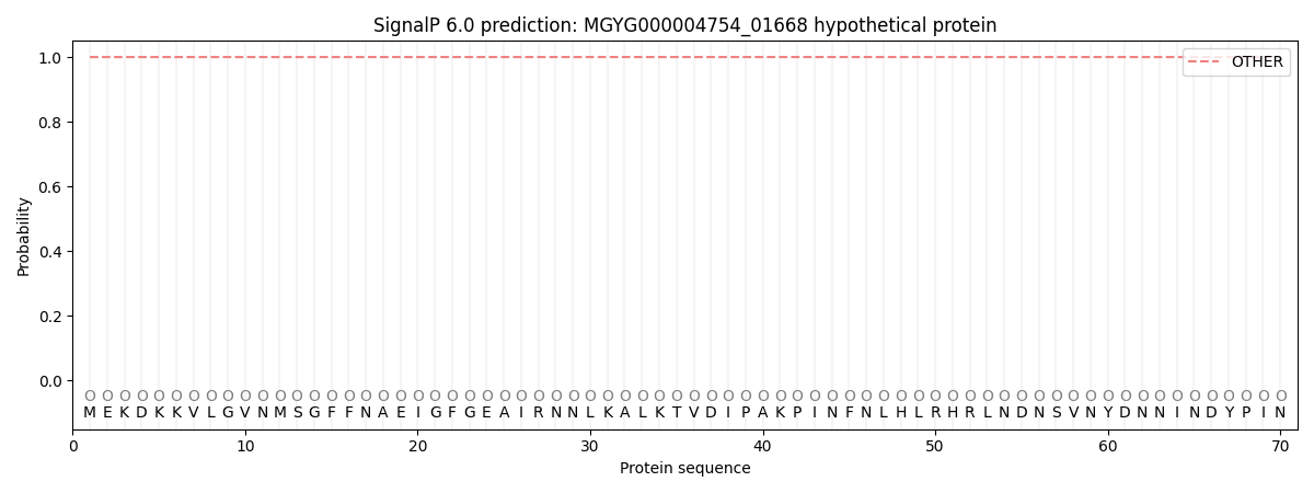
This protein is predicted as OTHER
| Other | SP_Sec_SPI | LIPO_Sec_SPII | TAT_Tat_SPI | TATLIP_Sec_SPII | PILIN_Sec_SPIII |
|---|---|---|---|---|---|
| 1.000062 | 0.000000 | 0.000000 | 0.000000 | 0.000000 | 0.000000 |