You are browsing environment: HUMAN GUT
CAZyme Information: MGYG000004864_02376
You are here: Home > Sequence: MGYG000004864_02376
Basic Information |
Genomic context |
Full Sequence |
Enzyme annotations |
CAZy signature domains |
CDD domains |
CAZyme hits |
PDB hits |
Swiss-Prot hits |
SignalP and Lipop annotations |
TMHMM annotations
Basic Information help
| Species | ||||||||||||
|---|---|---|---|---|---|---|---|---|---|---|---|---|
| Lineage | Bacteria; Desulfobacterota; Desulfovibrionia; Desulfovibrionales; Desulfovibrionaceae; Desulfovibrio; | |||||||||||
| CAZyme ID | MGYG000004864_02376 | |||||||||||
| CAZy Family | GT51 | |||||||||||
| CAZyme Description | Penicillin-binding protein 1C | |||||||||||
| CAZyme Property |
|
|||||||||||
| Genome Property |
|
|||||||||||
| Gene Location | Start: 147; End: 1538 Strand: + | |||||||||||
CDD Domains download full data without filtering help
| Cdd ID | Domain | E-Value | qStart | qEnd | sStart | sEnd | Domain Description |
|---|---|---|---|---|---|---|---|
| TIGR02073 | PBP_1c | 5.77e-143 | 1 | 459 | 285 | 726 | penicillin-binding protein 1C. This subfamily of the penicillin binding proteins includes the member from E. coli designated penicillin-binding protein 1C. Members have both transglycosylase and transpeptidase domains and are involved in forming cross-links in the late stages of peptidoglycan biosynthesis. All members of this subfamily are presumed to have the same basic function. [Cell envelope, Biosynthesis and degradation of murein sacculus and peptidoglycan] |
| COG4953 | PbpC | 1.93e-129 | 1 | 459 | 306 | 731 | Membrane carboxypeptidase/penicillin-binding protein PbpC [Cell wall/membrane/envelope biogenesis]. |
| PRK11240 | PRK11240 | 9.85e-58 | 1 | 450 | 309 | 762 | penicillin-binding protein 1C; Provisional |
| COG0744 | MrcB | 4.41e-47 | 2 | 256 | 339 | 596 | Membrane carboxypeptidase (penicillin-binding protein) [Cell wall/membrane/envelope biogenesis]. |
| TIGR02074 | PBP_1a_fam | 7.72e-46 | 2 | 264 | 268 | 531 | penicillin-binding protein, 1A family. Bacterial that synthesize a cell wall of peptidoglycan (murein) generally have several transglycosylases and transpeptidases for the task. This family consists of bifunctional transglycosylase/transpeptidase penicillin-binding proteins (PBP). In the Proteobacteria, this family includes PBP 1A but not the paralogous PBP 1B (TIGR02071). This family also includes related proteins, often designated PBP 1A, from other bacterial lineages. [Cell envelope, Biosynthesis and degradation of murein sacculus and peptidoglycan] |
CAZyme Hits help
| Hit ID | E-Value | Query Start | Query End | Hit Start | Hit End |
|---|---|---|---|---|---|
| SPD36822.1 | 8.00e-313 | 1 | 461 | 310 | 770 |
| ATD81196.1 | 8.00e-313 | 1 | 461 | 310 | 770 |
| VZH32198.1 | 1.23e-269 | 1 | 460 | 354 | 813 |
| QTO41888.1 | 8.68e-232 | 1 | 460 | 310 | 768 |
| QCC84882.1 | 2.71e-231 | 1 | 460 | 334 | 792 |
PDB Hits download full data without filtering help
| Hit ID | E-Value | Query Start | Query End | Hit Start | Hit End | Description |
|---|---|---|---|---|---|---|
| 4OON_A | 3.73e-14 | 3 | 249 | 408 | 715 | Crystalstructure of PBP1a in complex with compound 17 ((4Z,8S,11E,14S)-5-(2-amino-1,3-thiazol-4-yl)-14-(5,6-dihydroxy-1,3-dioxo-1,3-dihydro-2H-isoindol-2-yl)-8-formyl-2-methyl-6-oxo-3,10-dioxa-4,7,11-triazatetradeca-4,11-diene-2,12,14-tricarboxylic acid) [Pseudomonas aeruginosa PAO1] |
| 3UDF_A | 8.29e-14 | 3 | 249 | 400 | 708 | ChainA, Penicillin-binding protein 1a [Acinetobacter baumannii],3UDF_B Chain B, Penicillin-binding protein 1a [Acinetobacter baumannii],3UDI_A Chain A, Penicillin-binding protein 1a [Acinetobacter baumannii],3UDI_B Chain B, Penicillin-binding protein 1a [Acinetobacter baumannii],3UDX_A Chain A, Penicillin-binding protein 1a [Acinetobacter baumannii],3UDX_B Chain B, Penicillin-binding protein 1a [Acinetobacter baumannii],3UE0_A Chain A, Penicillin-binding protein 1a [Acinetobacter baumannii],3UE0_B Chain B, Penicillin-binding protein 1a [Acinetobacter baumannii],3UE1_A Chain A, Penicillin-binding protein 1a [Acinetobacter baumannii],3UE1_B Chain B, Penicillin-binding protein 1a [Acinetobacter baumannii] |
| 2FFF_B | 5.01e-12 | 27 | 261 | 119 | 373 | OpenForm of a Class A Transpeptidase Domain [Streptococcus pneumoniae] |
| 2JCH_A | 5.12e-12 | 27 | 261 | 127 | 381 | Structuraland mechanistic basis of penicillin binding protein inhibition by lactivicins [Streptococcus pneumoniae R6] |
| 2BG1_A | 5.55e-12 | 27 | 261 | 158 | 412 | Activesite restructuring regulates ligand recognition in classA Penicillin-binding proteins (PBPs) [Streptococcus pneumoniae R6],2XD5_A Structural insights into the catalytic mechanism and the role of Streptococcus pneumoniae PBP1b [Streptococcus pneumoniae R6],2XD5_B Structural insights into the catalytic mechanism and the role of Streptococcus pneumoniae PBP1b [Streptococcus pneumoniae R6] |
Swiss-Prot Hits download full data without filtering help
| Hit ID | E-Value | Query Start | Query End | Hit Start | Hit End | Description |
|---|---|---|---|---|---|---|
| P76577 | 1.09e-48 | 1 | 451 | 311 | 761 | Penicillin-binding protein 1C OS=Escherichia coli (strain K12) OX=83333 GN=pbpC PE=1 SV=1 |
| O87579 | 3.19e-19 | 3 | 261 | 435 | 710 | Penicillin-binding protein 1A OS=Neisseria lactamica OX=486 GN=mrcA PE=3 SV=1 |
| O86088 | 3.19e-19 | 3 | 254 | 435 | 703 | Penicillin-binding protein 1A OS=Neisseria cinerea OX=483 GN=mrcA PE=3 SV=1 |
| P0A0Z5 | 4.24e-19 | 3 | 254 | 435 | 703 | Penicillin-binding protein 1A OS=Neisseria meningitidis serogroup A / serotype 4A (strain DSM 15465 / Z2491) OX=122587 GN=mrcA PE=3 SV=1 |
| P0A0Z6 | 4.24e-19 | 3 | 254 | 435 | 703 | Penicillin-binding protein 1A OS=Neisseria meningitidis serogroup B (strain MC58) OX=122586 GN=mrcA PE=3 SV=1 |
SignalP and Lipop Annotations help
This protein is predicted as OTHER
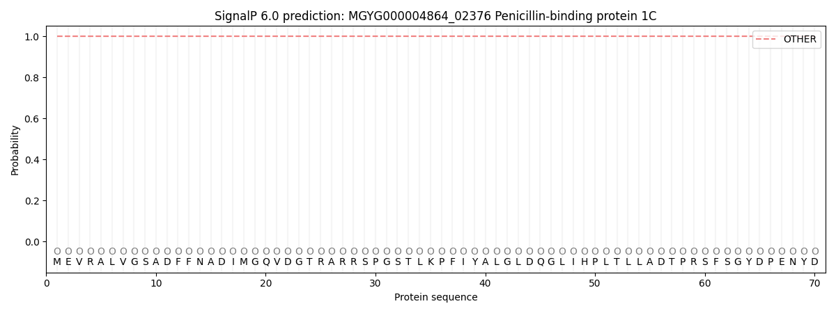
| Other | SP_Sec_SPI | LIPO_Sec_SPII | TAT_Tat_SPI | TATLIP_Sec_SPII | PILIN_Sec_SPIII |
|---|---|---|---|---|---|
| 1.000039 | 0.000000 | 0.000000 | 0.000000 | 0.000000 | 0.000000 |